Summary

与碘酸雪夫染色检测糖原的外周血单个核细胞

Published: December 23, 2014
doi:

Summary

Periodic acid Schiff staining is a technique that visualizes the polysaccharide content of tissues. This article demonstrates periodic acid Schiff staining protocol adapted for use on peripheral blood mononuclear cells purified from human venous blood. Such samples are enriched for lymphocytes and other white blood cells of the immune system.

Abstract

Periodic acid Schiff (PAS) staining is an immunohistochemical technique used on muscle biopsies and as a diagnostic tool for blood samples. Polysaccharides such as glycogen, glycoproteins, and glycolipids stain bright magenta making it easy to enumerate positive and negative cells within the tissue. In muscle cells PAS staining is used to determine the glycogen content in different types of muscle cells, while in blood cell samples PAS staining has been explored as a diagnostic tool for a variety of conditions. Blood contains a proportion of white blood cells that belong to the immune system. The notion that cells of the immune system possess glycogen and use it as an energy source has not been widely explored. Here, we describe an adapted version of the PAS staining protocol that can be applied on peripheral blood mononuclear immune cells from human venous blood. Small cells with PAS-positive granules and larger cells with diffuse PAS staining were observed. Treatment of samples with amylase abrogates these patterns confirming the specificity of the stain. An alternate technique based on enzymatic digestion confirmed the presence and amount of glycogen in the samples. This protocol is useful for hematologists or immunologists studying polysaccharide content in blood-derived lymphocytes.

Introduction

碘酸希夫(PAS)染色是一种免疫组织化学技术,被广泛用于肌肉研究和诊断。它也被用来作为对血样的诊断工具。该技术的工作原理是将高碘酸溶液的样品,其氧化,与无色希夫氏试剂,从而产生了深刻的品红产物反应的多糖产生醛基内单元。这个过程的步骤显示在图1的染色变为与多糖品红,包括糖原,糖蛋白,糖脂,粘蛋白,或其它分子与多糖部分任何东西。

PAS染色常常用于测量肌肉纤维糖原水平。肌肉组织切片是理想的,因为他们牢牢地固定在滑板和承受多次洗涤和染色步骤的技术。糖原是最存在于快肌II型肌纤维,它具有高的需求快速ATP生产要求糖原的最高性能1,2。糖原是葡萄糖的支链聚合物,可以通过对糖原磷酸酶的作用被分解成游离葡萄糖。在休息和营养自足倍,糖原通过糖原的过程中补充,而在营养不足或高能量需求的时间;糖原是由糖原分解分解成葡萄糖。从早在1950年的临床科学家们探索的血样PAS染色法来分析各种疾病3-7糖原含量。例如,在蓬珀病-一个真实糖原贮积无病白细胞积累大量糖原,从健康对照8显著不同的。

此视频文章演示了PAS染色的外周血单个核细胞(PBMC)从健康人的静脉血样使用改编版。 PBM铯包含大多淋巴细胞T淋巴细胞和B淋巴细胞的家庭,以及其他免疫细胞如自然杀伤细胞和单核细胞。第一纯化步骤除去红细胞,嗜中性粒细胞,和其它粒细胞。该技术提供了对淋巴细胞允许PAS阳性细胞的更健壮的枚举,相比使用全血涂片的浓缩比例的数据。

图1
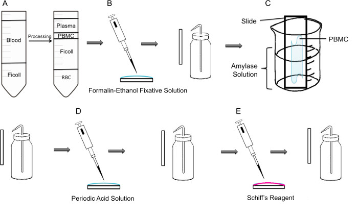
图1:第一步通过PBMC PAS染色步骤方法(一)首先,PBMC的隔离是通过聚蔗糖梯度实现的,左边的面板显示离心前的准备,右侧面板离心后显示它那里的棕黄色大衣含PBMC在管的中心观察到。(B)中分离的PBMC中使用福尔马林固定液乙醇SOLU固定到滑动化。滑动轻轻漂洗由塑料洗瓶蒸馏水。(C)的滑动,然后放置在填充有淀粉酶溶液的100毫升烧杯中的一半,这将溶解糖原。滑动轻轻漂洗。(D)将载玻片用高碘酸溶液中,其中糖的氧化发生处理。幻灯片轻轻冲洗;这将除去多余的高碘酸并停止氧化步骤。(E)当希夫试剂被添加到幻灯片,它将与在氧化步骤中创建的醛反应。然后,该无色试剂将导致深红色深红色产物。载玻片轻轻冲洗以除去过量的希夫试剂。

Protocol

研究与人类血液样本被批准Concordia大学伦理审查委员会,证书编号10000618.批准了康考迪亚大学伦理审查委员会,证书编号2010BERG对小鼠肌肉的工作。 从全血1 PBMC隔离注:执行此过程中使用无菌技术和制造商消毒设备生物安全柜。 小心倒入10-15毫升的肝素化(抗凝血剂)血液收集管的全血到50ml无菌锥形管中。对于轻微的血液外溢,使用清洁纸巾擦拭…

Representative Results

为了验证所用试剂和基本技术,PAS染色物根据制造商的对小鼠比目鱼肌节的指令进行的。染色做同一天的牺牲,并在染色的最后步骤,将切片固定二甲苯。比目鱼肌已知含有〜35%的糖原阳性细胞9。染色的肌肉细胞中显示两个不同的PAS阳性特性-点状颗粒的细胞内,并且连续线demarking细胞膜( 图2A)。点状颗粒的存在是与糖原储存一致,并且被用来定义一个阳性肌细胞。用淀?…

Discussion

这个视频文章的关键步骤是在洗涤和淀粉酶处理的细胞。而洗涤载玻片,关键步骤是使用塑料挤压瓶洗涤并让水通过幻灯片上的样品轻轻地运行,并没有直接瞄准到样品。即使是最轻微的直接供水压力会导致细胞脱落的幻灯片。另一个关键步骤是使用相同的幻灯片±淀粉酶的条件。后的PBMC附着到滑动,滑动小心地放入烧杯所以只有一半的涂片的暴露于淀粉酶溶液。此步骤提供了一个强大的控制,?…

Declarações

The authors have nothing to disclose.

Acknowledgements

This research was supported by a grant from the NSERC Discovery program grant number RGPIN 418522-2013. We thank R. Kilgour for helpful discussions, and Katelin Gresty and Dr. A. Berghdal for providing the mouse muscle sections.

Materials

Periodic Acid Shiff Kit Sigma-Aldrich 395B Bring to room temperature prior to use. Materials in this kit are toxic and harmful. Use caution 
http://www.sigmaaldrich.com/catalog/product/sigma/395b?lang=en&region=CA
α-Amylase from porcine pancreas Sigma-Aldrich A3176 http://www.sigmaaldrich.com/catalog/product/sigma/a3176?lang=en&region=CA
Binocular Microscope  Carl Zeiss Microscopy Axio Lab A0
Glycogen Assay Kit Sigma-Aldrich MAK016 http://www.sigmaaldrich.com/catalog/product/sigma/mak016?lang=en&region=CA
Ficoll-Paque PLUS VWR, GE Healthcare 17-1440-02 Nonionic synthetic polymer of sucrose
https://us.vwr.com/store/catalog/product.jsp?product_id=4779441
Centrifuge For PBMC isolation, swing buckets were used 

Referências

  1. Rich, P. R. The molecular machinery of keilin’s respiratory chain. Biochem. Soc. Trans. 31 (Pt 6), 1095-1105 (2003).
  2. Peter, J. B., Barnard, R. J., Edgerton, V. R., Gillespie, C. A., Stempel, K. E. Metabolic profiles of three fiber types of skeletal muscle in guinea pigs and rabbits). Bioquímica. 11 (14), 2627-2633 (1972).
  3. Jones, R. V., Goffi, G. P., Hutt, M. S. R. Lymphocyte glycogen content in various disease. J. Clin. Pathol. 15 (1), 36-39 (1962).
  4. Scott, R. B. Glycogen in human peripheral blood leukocytes. I. characteristics of the synthesis and turnover of glycogen in vitro. J. Clin. Invest. 47 (2), 344-352 (1968).
  5. Fedele, D., et al. positive index of lymphocytes and metabolic control in insulin-treated and type II diabetes mellitus. Diabete Metab. 9 (3), 188-192 (1983).
  6. Brelińska-Peczalska, R., Mackiewicz, S. Cytochemical studies of peripheral blood granulocytes and lymphocytes in patients with systemic lupus erythematosus. Pol.Med.Sci.Hist.Bull. 15 (2), 231-234 (1976).
  7. Yunis, A. A., Arimura, G. K. Enzymes of glycogen metabolism in white blood cells. I. glycogen phosphorylase in normal and leukemic human leukocytes. Cancer, Res. 24, 489-492 (1964).
  8. Hagemans, M. L., et al. PAS-positive lymphocyte vacuoles can be used as diagnostic screening test for pompe disease. J. Inherit. Metab. Dis. 33 (2), 133-139 (2010).
  9. Totsuka, Y., et al. Physical performance and soleus muscle fiber composition in wild-derived and laboratory inbred mouse strains. 95 (2), 720-727 (2003).
  10. Murat, J. C., Serfaty, A. Simple enzymatic determination of polysaccharide (glycogen) content of animal tissues. Clin. Chem. 20 (12), 1576-1577 (1974).
  11. Arrizabalaga, O., Lacerda, H. M., Zubiaga, A. M., Zugaza, J. L. Rac1 protein regulates glycogen phosphorylase activation and controls interleukin (IL)-2-dependent T cell proliferation. J. Biol. Chem. 287 (15), 11878-11890 (2012).
  12. Pelletier, J., G, J., Mazure, N. M. Biochemical titration of glycogen in vitro. J.Vis.Exp. (81), (2013).
  13. Roach, P. J., Depaoli-Roach, A. A., Hurley, T. D. Tagliabracci V.S. Glycogen and its metabolism: Some new developments and old themes. Biochem.J. 441 (3), 763-787 (2012).
  14. Salmoral, E. M., Tolmasky, D. S., Krisman, C. R. Evidence for the presence of glycogen in rat thymus. Cell Mol.Biol. 36 (2), 163-174 (1990).
  15. Darlington, P. J., et al. Diminished Th17 (not Th1) responses underlie multiple sclerosis disease abrogation after hematopoietic stem cell transplantation. Ann.Neurol. 73 (3), 341-354 (2013).
check_url/pt/52199?article_type=t

Play Video

Citar este artigo
Tabatabaei Shafiei, M., Carvajal Gonczi, C. M., Rahman, M. S., East, A., François, J., Darlington, P. J. Detecting Glycogen in Peripheral Blood Mononuclear Cells with Periodic Acid Schiff Staining. J. Vis. Exp. (94), e52199, doi:10.3791/52199 (2014).

View Video