Summary

마우스 폐 전 절제술 및 보철물 삽입을 통한 내시경 표면 측정을위한 표준화 된 방법

Published: July 26, 2017
doi:

Summary

내부 폐 표면적 (ISA)은 폐 질환 및 부상 유발 폐포 재생에서 폐 형태 및 생리학을 평가하는 데 중요한 기준입니다. 폐 절제술 및 인공 삽입물 마우스 모델에서 ISA의 측정 편향을 최소화 할 수있는 표준화 된 방법을 설명합니다.

Abstract

Pulmonary morphology, physiology, and respiratory functions change in both physiological and pathological conditions. Internal lung surface area (ISA), representing the gas-exchange capacity of the lung, is a critical criterion to assess respiratory function. However, observer bias can significantly influence measured values for lung morphological parameters. The protocol that we describe here minimizes variations during measurements of two morphological parameters used for ISA calculation: internal lung volume (ILV) and mean linear intercept (MLI). Using ISA as a morphometric and functional parameter to determine the outcome of alveolar regeneration in both pneumonectomy (PNX) and prosthesis implantation mouse models, we found that the increased ISA following PNX treatment was significantly blocked by implantation of a prosthesis into the thoracic cavity1. The ability to accurately quantify ISA is not only expected to improve the reliability and reproducibility of lung function studies in injured-induced alveolar regeneration models, but also to promote mechanistic discoveries of multiple pulmonary diseases.

Introduction

폐의 기본 기능은 혈관과 대기 사이의 산소와 이산화탄소의 교환입니다. 기관지 폐 이형성증 (BPD), 만성 폐색 성 폐 질환 (COPD) 및 급성 호흡기 감염과 같은 폐 질환은 ISA 2 를 감소시킵니다. 폐 질환을 연구하는 연구자들은 MLI, ILV, 기체 교환 단위 수, ISA 및 폐 조직 적합성 2 , 3을 포함하여 폐의 형태 학적 변화를 평가하기위한 몇 가지 정량적 방법을 개발했습니다. Weibel 등의 선구자 연구 4 및 Duguid et al. 5는 함께 ISA가 인간의 폐에서 폐 가스 교환 용량의 직접 척도로 사용될 수 있음을 입증했으며 기종의 심각성을 판단하는 기준으로 사용할 수있다. 지난 5 년 동안 발표 된 여러 연구에서 폐 형태 학적 매개 변수 ( 예 : </em> ISA 및 MLI)가 개발 중에 6 및 부상 PNX 1 (7)로부터 복구시 쥐의 폐에 형태 학적 및 기능적 변화를 평가한다. ISA는 식 1 8 , 9를 사용하여 계산됩니다.

방정식

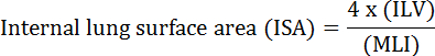
, ILV 내부 폐 용적이며 MLI는 말초 폐 공역 10 크기를 나타내는 매개 변수이다.

하나 이상의 폐엽의 외과 적 제거 인 PNX는 인간 11 , 마우스 1 , 개 12 , 쥐 13 및 토끼 14 , 15를 비롯한 많은 종에서 폐포 재생을 유도하는 것으로 널리보고되었습니다. 스터드PNX 후 14 일째 생쥐의 폐는 기존 폐포의 팽창과 폐포의 새로운 형성 모두 남아있는 폐 조직에서 ISA, ILV 및 폐포의 수를 회복시키는 데 기여한다고보고했다 1 . 우리와 다른 사람들은 스폰지, 왁스 또는 주문형 보철물과 같은 재료를 PNX ( , 보철물 삽입) 후 빈 흉부 공동에 삽입하면 폐포 재생을 손상시키는 것으로 나타났습니다. 기계적 힘은 폐포 재생을 시작하는 가장 중요한 요인 중 하나로 작용한다고 확고하게 확립되었습니다 1 , 16 , 17 . 이러한 연구는 PNX 치료 및 인공 삽입물 폐에서 ISA 값을 사용하여 폐포 재생을 정량적으로 평가하는 기준으로 사용하는 것이 효과적이라는 것을 강조했습니다.

옵서버 바이어스는 측정 된 VA에 유의 한 영향을 미치는 것으로 알려져 있습니다.폐 형태 학적 매개 변수 ( 예 : MIL 및 ILV)에 대한 정보. 표준화 된 프로토콜은 ISA 계산에 사용 된 두 가지 매개 변수 인 ILV와 MLI를 결정할 때이 편향을 없애기 위해 사용할 수 있습니다. 여기서는 이러한 폐 매개 변수를 측정하기위한 매우 상세한 표준화 된 프로토콜을 제공합니다. 중요하게도, ISA를 정확히 정량화하는 능력은 손상 유발 폐포 재생 모델에서 폐 기능 연구의 신뢰성과 재현성을 향상시키고 다중 폐 질환에서 기계적 발견을 촉진해야한다고 약속한다.

Protocol

이 프로토콜에 사용 된 모든 절차는 국립 생물 과학 연구소의 실험실 동물의 관리 및 사용에 관한 지침의 권장 사항에 따라 수행되었습니다. 8 주된 CD-1 수컷 마우스를 실험이 수행 될 때까지 특정 병원균이없는 (SPF) 시설에 수용했다. 수술은 완전히 마취 된 마우스를 사용하여 수행되었습니다 ( 즉 , 발가락 핀치 반응없이). 수술 후 쥐는 충분한 음식과 담수가있는 따뜻하고 습기가 많은 방?…

Representative Results

우리는 여기서 PNX 치료군과 인공 삽입물 (인공 삽입물)을 사용한 실험을 수행했습니다. 이러한 그룹은 이전에 발표 된 연구 그룹의 연구에서 사용한 그룹과 동일합니다 14 . 마우스 PNX 및 보철물 삽입 절차는 그림 2에 나와 있습니다. 8 주 된 CD-1 수컷 마우스가 수술과 정량화에 사용…

Discussion

이 프로토콜에서는 마우스 왼쪽 폐 PNX 및 보철물 삽입 후 폐 매개 변수 측정에 대한 자세한 설명을 제공합니다. ISA는 현재 많은 폐 질환 및 부상 유발 폐포 재생에서 호흡 기능 평가의 주요 척도로 간주됩니다. 그러나 폐 연구 공동체가 ISA의 유용성에 관해서는 유용한 척도로 동의하고 있지만 현재까지는 ISA를 계산하는 데 사용 된 두 가지 매개 변수 인 ILV 및 MLI 측정의 표준화에 대해서는 거의 고?…

Declarações

The authors have nothing to disclose.

Acknowledgements

저자는 북경 생물 과학 연구소 (Beijing Institute of Biological Sciences)에 도움을 청한다. 이 연구는 베이징 시립 자연 과학 재단 (No. Z17110200040000)의 지원을 받았다.

Materials

Low cost cautery kit Fine Science Tools 18010-00
Noyes scissors Fine Science Tools 15012-12
Standard pattern forceps Fine Science Tools 11000-12
Castroviejo Micro Needle Holders Fine Science Tools 12060-01
Vessel clips Fine Science Tools 18374-44
I. V. Cannula-20 gauge Jinhuan Medical Product Co., LTD. 29P0601
Surgical suture Jinhuan Medical Product Co., LTD. F602
Mouse intubation platform Penn-Century, Inc Model MIP
Small Animal Laryngoscope Penn-Century, Inc Model LS-2-M
TOPO Small Animal Ventilator Kent Scientific RSP1006-05L
Thermal pad Stuart equipment SBH130D
Pentobarbital sodium salt Sigma P3761
Heparin sodium salt Sigma H3393
Hematoxylin Solution Sigma GHS132
Eosin Y solution, alcoholic Sigma HT110116
10 ml Pipette Thermo Scientific 170356
Paraformaldehyde Sigma P6148
O.C.T Compound Tissue-Tek 4583
cryosection machine Leica CM1950
Disposable Base Molds Fisher HealthCare 22-363-553
18 gauge needle Becton Dickinson 305199
Povidone iodine Fisher Scientific 19-027132
70% ethanol Fisher Scientific BP82011
Infusion sets for single use Weigao SFDA 2012 3661704
Phosphate buffered saline Gibco 10010023
Tapes 3M Scotch 8915
Cotton pad Vinda Dr.P
Silicone prosthesis Custom made
Brightfield microscope Olympus VS120
Ruler tool Adobe Photoshop

Referências

  1. Liu, Z., et al. MAPK-Mediated YAP Activation Controls Mechanical-Tension-Induced Pulmonary Alveolar Regeneration. Cell Rep. 16 (7), 1810-1819 (2016).
  2. Thurlbeck, W. M. Internal surface area and other measurements in emphysema. Thorax. 22 (6), 483-496 (1967).
  3. Knudsen, L., Weibel, E. R., Gundersen, H. J. G., Weinstein, F. V., Ochs, M. Assessment of air space size characteristics by intercept (chord) measurement: an accurate and efficient stereological approach. J Appl Physiol. 108 (2), 412-421 (2010).
  4. Weibel, E. R. . Morphometry of the Human Lung. , (1963).
  5. Duguid, J. B., Young, A., Cauna, D., Lambert, M. W. The internal surface area of the lung in emphysema. J Pathol Bacteriol. 88, 405-421 (1964).
  6. Branchfield, K., et al. Pulmonary neuroendocrine cells function as airway sensors to control lung immune response. Science. 351 (6274), 707-710 (2016).
  7. Ding, B. -. S., et al. Endothelial-derived angiocrine signals induce and sustain regenerative lung alveolarization. Cell. 147 (3), 539-553 (2011).
  8. Dunnill, M. S. Quantitative methods in the study of pulmonary pathology. Thorax. 17 (4), 320-328 (1962).
  9. Weibel, E. R., Gomez, M. Architecture of the human lung. Use of quantitative methods establishes fundamental relations between size and number of lung structures. Science. 137 (3530), 577-585 (1962).
  10. Thurlbeck, W. M. The internal surface area of nonemphysematous lungs. Am Rev Respir Dis. 95 (5), 765-773 (1967).
  11. Butler, J. P., et al. Evidence for adult lung growth in humans. N Engl J Med. 367 (16), 244-247 (2012).
  12. Hsia, C. C. W., Herazo, L. F., Fryder-Doffey, F., Weibel, E. R. Compensatory lung growth occurs in adult dogs after right pneumonectomy. J Clin Invest. 94 (1), 405-412 (1994).
  13. Thurlbeck, S. W. M. Pneumonectomy in Rats at Various Ages. Am Rev Respir Dis. 120 (5), 1125-1136 (1979).
  14. Cagle, P. T., Langston, C., Thurlbeck, W. M. The Effect of Age on Postpneumonectomy Growth in Rabbits. Pediatr Pulmonol. 5 (2), 92-95 (1988).
  15. Langston, C., et al. Alveolar multiplication in the contralateral lung after unilateral pneumonectomy in the rabbit. Am Rev Respir Dis. 115 (1), 7-13 (1977).
  16. Cohn, R. Factors Affecting The Postnatal Growth of The Lung. Anatomical Record. 75 (2), 195-205 (1939).
  17. Hsia, C. C., Wu, E. Y., Wagner, E., Weibel, E. R. Preventing mediastinal shift after pneumonectomy impairs regenerative alveolar tissue growth. Am J Physiol Lung Cell Mol Physiol. 281 (5), L1279-L1287 (2001).
  18. Das, S., MacDonald, K., Chang, H. -. Y. S., Mitzner, W. A simple method of mouse lung intubation. J Vis Exp. (73), e50318 (2013).
  19. Liu, S., Cimprich, J., Varisco, B. M. Mouse pneumonectomy model of compensatory lung growth. J Vis Exp. (94), (2014).
  20. Silva, M. F. R., Zin, W. A., Saldiva, P. H. N. Airspace configuration at different transpulmonary pressures in normal and paraquat-induced lung injury in rats. Am J Respir Crit Care Med. 158 (4), 1230-1234 (1998).
  21. Yilmaz, C., et al. Noninvasive quantification of heterogeneous lung growth following extensive lung resection by high-resolution computed tomography. J Appl Physiol. 107 (5), 1569-1578 (2009).
  22. Voswinckel, R., et al. Characterisation of post-pneumonectomy lung growth in adult mice. Eur Respir J. 24 (4), 524-532 (2004).
  23. Ravikumar, P., et al. Regional Lung Growth and Repair Regional lung growth following pneumonectomy assessed by computed tomography. J Appl Physiol. 97, 1567-1574 (2004).
  24. Gibney, B. C., et al. Detection of murine post-pneumonectomy lung regeneration by 18FDG PET imaging. EJNMMI Res. 2 (1), (2012).
  25. Muñoz-Barrutia, A., Ceresa, M., Artaechevarria, X., Montuenga, L. M., Ortiz-De-Solorzano, C. Quantification of lung damage in an elastase-induced mouse model of emphysema. Int J Biomed Imaging. 2012, (2012).
check_url/pt/56114?article_type=t

Play Video

Citar este artigo
Liu, Z., Fu, S., Tang, N. A Standardized Method for Measuring Internal Lung Surface Area via Mouse Pneumonectomy and Prosthesis Implantation. J. Vis. Exp. (125), e56114, doi:10.3791/56114 (2017).

View Video