Summary

鼠标离体肾技术

Published: November 17, 2016
doi:

Summary

鼠标离体灌注肾(MIPK)是用于灌注和1小时官能体外条件下保持一个小鼠肾脏的技术。的缓冲液和外科技术进行详细说明。

Abstract

鼠标离体灌注肾(MIPK)是用于灌注和1小时官能体外条件下保持一个小鼠肾脏的技术。这是研究分离的器官,并为许多创新的应用程序,可能会在未来成为可能,包括灌注脱细胞肾生物工程或抗排斥或基因组编辑药物在高剂量黄金肾脏的管理生理学的一个先决条件用于移植。在灌注时,肾脏可以被操纵,肾功能可以评估,和给药的各种药物。手术后,肾脏可以移植或用于分子生物学,生物化学分析,或显微处理。

本文介绍了灌流,需要鼠标肾脏的体外灌注的手术技术。灌注装置的细节给出并且呈现表示V数据肾脏的制剂iability:肾血流量,血管阻力,和尿的数据作为不同肾段作为形态学读数,和不同肾段作为分子读出的转运蛋白的蛋白质印迹的官能,透射电子显微镜照片。

Introduction

器官的隔离灌注一直生理学家中不断努力的主题几十年1。该技术使器官的功能,而没有全身的影响,如血压,激素,或神经,进行研究。卡尔·爱德华Loebell被认为是第一个所描述的一个孤立肾灌注成功,于1849年2。此后,灌注设备发生了显著细化。弗雷和格鲁伯引入了氧合搏动泵人工肺的连续灌流2。虽然早期的研究人员重点研究大型哺乳动物-即,猪2和狗3使用大鼠肾脏的-the第一份报告,魏斯等人的肾脏 ,是小型哺乳动物器官灌注4研究的一个里程碑。 Schurek 等。报道添加哺乳动物红细胞到灌流如果有足够的肾小管的必要性氧合要达到5。临界长期实验是由相同的研究组6引进缓冲器的连续透析。在2003年,Schweda 等。是第一个报告功能的鼠标离体灌注肾(MIPK)7,后来被Rahgozar 精制而成 18和林德尔等。 14。

虽然在技术上不是孤立肾灌注大鼠更具挑战性,使用MIPK负有能够使用各种各样基因改造的小鼠中的优势。本文介绍了作者的方法的详细信息隔离灌注小鼠肾1小时。该方法允许对肾流量,血管阻力,激素释放,血气分析,尿液分析,以及药物的应用程序的连续评估。以下的方法,肾脏可以用于分子和生化分析进行处理,是固定的显微镜,或移植到受体小鼠( 图1)。

图1
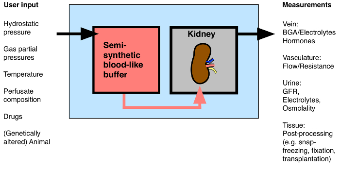
图1:可能的输入/输出到离体肾的概述。 BGA:血气分析。 请点击此处查看该图的放大版本。

这种技术可能会得到更多的关注,未来数年,多创新应用正在与长期常温肾脏血流灌注的曙光移植前的讨论(含或不含抗排斥或基因组编辑药物的应用)8,9,10 11,从脱细胞支架12整个肾脏的生物工程,剂量和高剂量荧光染料对多光子成像13应用</sup>。这也是与研究急性肾损伤14中特定基因的作用的理想模型。

一个一步一步的协议,是考虑到让其他实验室成功地执行孤立的小鼠肾脏灌注。首先,组合物和制剂的缓冲液中被指定。然后,手术中详细描述和示出的关键步骤。第三,数据呈现的是,用于展示成功制备的:肾血流量,血管阻力,肾小球滤过率和分数电解质排泄-所有灌注肾脏的不同肾段的形态的生存能力和透射电子显微照片的功能性测量灌注后的1小时固定。

Protocol

所有涉及这个手稿中描述动物的程序进行了根据瑞士法律和瑞士苏黎世州的兽医行政管理部门批准。 1.缓冲液配制制备溶液1 – 4和抗利尿激素(ADH)溶液( 表1)。 制备透析缓冲液( 表1)。 注意:这是用作灌注在透析缓冲的缓冲器。以后,红细胞将在这个缓冲液稀释以形成最终灌洗液。 红细胞制剂。 稀释250毫升人红…

Representative Results

与所描述的方法,分离小鼠肾可为至少1小时保持存活。我们后官能(肾血流量和血管阻力,静脉流出,肾小球滤过率,尿分数Na +和K +排泄血气分析和尿液摩尔渗透压浓度)和形态(透射电子连续灌注的1小时测试的组织活力显微镜,TEM)的方法,在野生型C57BL / 6小鼠四个肾脏。此外,进行了不同的小管的根尖标记蛋白的蛋白质印迹,隔离灌注肾脏比较同一…

Discussion

离体灌注肾脏鼠标是用于在受控环境中的体外研究肾功能1小时,架桥在完整的动物体内实验,这可以通过许多全身性因素的影响,是有缺陷的之间的间隙的一个工具,并在体外实验中分离的肾段或培养细胞,这必然忽视完整器官结构对功能的影响。还有就是,以作者的知识,没有替代技术,用以执行这一特定的任务。对肾组织的生化研究,然而,可以在使用肾脏切片17</sup…

Disclosures

The authors have nothing to disclose.

Acknowledgements

The authors would like to thank Hans-Joachim Schurek for invaluable scientific advice. The authors would like to thank Monique Carrel and Michèle Heidemeyer for excellent technical assistance, David Penton Ribas and Nourdine Faresse for a critical reading of the manuscript and Carsten Wagner and Jürg Biber for the NaPi-2a antibody. This work was supported by the Swiss National Centre for Competence in Research “Kidney.CH” and by a project grant (310030_143929/1) from the Swiss National Science Foundation.

Materials

Perfusion Circuit:
Moist chamber 834/8 Harvard Apparatus/Hugo Sachs Elektronik GmbH 73-2901
Cannular with basket and side port Harvard Apparatus/Hugo Sachs Elektronik GmbH 73-2947
Thermostat TC120-ST5  Harvard Apparatus/Hugo Sachs Elektronik GmbH 73-4544
ISM 827/230V Roller Pump Reglo Analogue Harvard Apparatus/Hugo Sachs Elektronik GmbH 73-0114
Reservoir jacketed for buffer solution 1L Harvard Apparatus/Hugo Sachs Elektronik GmbH 73-3438
Reservoir jacketed for buffer solution 0.5L Harvard Apparatus/Hugo Sachs Elektronik GmbH 73-3436
Pressure Transducer APT300  Harvard Apparatus/Hugo Sachs Elektronik GmbH 73-3862
TAM-D Plugsys Transducer Harvard Apparatus/Hugo Sachs Elektronik GmbH 73-1793
SCP Plugsys servo controller Harvard Apparatus/Hugo Sachs Elektronik GmbH 73-2806
Windkessel  Harvard Apparatus/Hugo Sachs Elektronik GmbH 73-3717
HSE-USB data acquisition Harvard Apparatus/Hugo Sachs Elektronik GmbH 73-3330
Low-Flux Dialysator Diacap Polysulfone B.Braun 7203525
PE-Tubing for aorta cannulation 1.19mm I.D. x 1.70mm O.D. Scientific Commodities Inc. BB31695-PE/8
Name Company Catalog Number Comments
Buffer reagents:
Aminoplasmal 10% B.Braun 134518064
Sodium pyruvate Sigma-Aldrich P2256-25G
L-Glutamic acid monosodium salt hydrate Sigma-Aldrich G1626-100G
L-(-)-Malic acid sodium salt Sigma-Aldrich M1125-25G
Sodium-L-Lactate Sigma-Aldrich L7022-10G
alpha-Ketoglutaric acid sodium salt Sigma-Aldrich K1875-25G
NaCl Sigma-Aldrich 31434-1KG-R
NaHCO3 Sigma-Aldrich S5761-5KG
KCl Sigma-Aldrich 60130-1KG
Urea Sigma-Aldrich U5378-500G
Creatinine Sigma-Aldrich C4255-10G
Ampicillin Roche 10835242001
MgCl2 * 6H2O Sigma-Aldrich M2393-500G
D-Glucose Sigma-Aldrich G8270-1KG
CaCl2 * 6H2 Riedel-de-Haën 12074
NaH2PO4 Sigma-Aldrich S9638-500G
Na2HPO4 Sigma-Aldrich S0876-500G
Antidiuretic Hormone dDAVP Sigma-Aldrich V2013-1MG
FITC-Inulin Sigma-Aldrich
Filter used for erythrocyte filtration Macherey-Nagel MN 615
BGA Analysis:
ABL 80 flex Radiometer Medical ApS
Electron Microscope:
Philips CM100 TEM FEI

References

  1. Carrel, A., Lindbergh, C. A. The culture of whole organs. Science (New York, N.Y.). 81 (2112), 621-623 (1935).
  2. Skutul, K. . Über Durchströmungsapparate Pflüger’s Archiv. 123 (4-6), 249-273 (1908).
  3. Nizet, A. The isolated perfused kidney: possibilities, limitations and results. Kidney Int. 7 (1), 1-11 (1975).
  4. Weiss, C., Passow, H., Rothstein, A. Autoregulation of flow in isolated rat kidney in the absence of red cells. Am J Physiol. 196 (5), 1115-1118 (1959).
  5. Schurek, H. J., Kriz, W. Morphologic and functional evidence for oxygen deficiency in the isolated perfused rat kidney. Lab Invest. 53 (2), 145-155 (1985).
  6. Stolte, H., Schurek, H. J., Alt, J. M. Glomerular albumin filtration: a comparison of micropuncture studies in the isolated perfused rat kidney with in vivo experimental conditions. Kidney Int. 16 (3), 377-384 (1979).
  7. Schweda, F., Wagner, C., Krämer, B. K., Schnermann, J., Kurtz, A. Preserved macula densa-dependent renin secretion in A1 adenosine receptor knockout mice. AJP Renal Physiol. 284 (4), F770-F777 (2003).
  8. Moers, C., Smits, J. M., et al. Machine perfusion or cold storage in deceased-donor kidney transplantation. N Engl J Med. 360 (1), 7-19 (2009).
  9. Nicholson, M. L., Hosgood, S. A. Renal transplantation after ex vivo normothermic perfusion: the first clinical study. Am J Transplant. 13 (5), 1246-1252 (2013).
  10. Worner, M., Poore, S., Tilkorn, D., Lokmic, Z., Penington, A. J. A low-cost, small volume circuit for autologous blood normothermic perfusion of rabbit organs. Art Org. 38 (4), 352-361 (2014).
  11. Kaths, J. M., Spetzler, V. N., et al. Normothermic Ex Vivo Kidney Perfusion for the Preservation of Kidney Grafts prior to Transplantation. J Vis Exp. (101), e52909 (2015).
  12. Song, J. J., Guyette, J. P., Gilpin, S. E., Gonzalez, G., Vacanti, J. P., Ott, H. C. Regeneration and experimental orthotopic transplantation of a bioengineered kidney. Nature Med. 19 (5), 646-651 (2013).
  13. Hall, A. M., Crawford, C., Unwin, R. J., Duchen, M. R., Peppiatt-Wildman, C. M. Multiphoton imaging of the functioning kidney. JASN. 22 (7), 1297-1304 (2011).
  14. Lindell, S. L., Williams, N., Brusilovsky, I., Mangino, M. J. Mouse IPK: A Powerful Tool to Partially Characterize Renal Reperfusion and Preservation Injury. Open Transplant J. 5, 15-22 (2011).
  15. Wagner, C., De Wit, C., Kurtz, L., Grünberger, C., Kurtz, A., Schweda, F. Connexin40 is essential for the pressure control of renin synthesis and secretion. Circ Res. 100 (4), 556-563 (2007).
  16. Czogalla, J., Vohra, T., Penton, D., Kirschmann, M., Craigie, E., Loffing, J. The mineralocorticoid receptor (MR) regulates ENaC but not NCC in mice with random MR deletion. Pflüger’s Archiv. , (2016).
  17. Poosti, F., et al. Precision-cut kidney slices (PCKS) to study development of renal fibrosis and efficacy of drug targeting ex vivo. Dis Model Mech. 8 (10), 1227-1236 (2015).
  18. Rahgozar, M., Guan, Z., Matthias, A., Gobé, G. C., Endre, Z. H. Angiotensin II facilitates autoregulation in the perfused mouse kidney: An optimized in vitro model for assessment of renal vascular and tubular function. Nephrology. 9 (5), 288-296 (2004).
check_url/54712?article_type=t

Play Video

Cite This Article
Czogalla, J., Schweda, F., Loffing, J. The Mouse Isolated Perfused Kidney Technique. J. Vis. Exp. (117), e54712, doi:10.3791/54712 (2016).

View Video