Summary

A Mass Spectrometry-Based Approach to Identify Phosphoprotein Phosphatases and their Interactors

Published: April 29, 2022
doi:

Summary

Here, we present a protocol for the enrichment of endogenous phosphoprotein phosphatases and their interacting proteins from cells and tissues and their identification and quantification by mass spectrometry-based proteomics.

Abstract

Most cellular processes are regulated by dynamic protein phosphorylation. More than three-quarters of proteins are phosphorylated, and phosphoprotein phosphatases (PPPs) coordinate over 90% of all cellular serine/threonine dephosphorylation. Deregulation of protein phosphorylation has been implicated in the pathophysiology of various diseases, including cancer and neurodegeneration. Despite their widespread activity, the molecular mechanisms controlling PPPs and those controlled by PPPs are poorly characterized. Here, a proteomic approach termed phosphatase inhibitor beads and mass spectrometry (PIB-MS) is described to identify and quantify PPPs, their posttranslational modifications, and their interactors in as little as 12 h using any cell line or tissue. PIB-MS utilizes a non-selective PPP inhibitor, microcystin-LR (MCLR), immobilized on sepharose beads to capture and enrich endogenous PPPs and their associated proteins (termed the PPPome). This method does not require the exogenous expression of tagged versions of PPPs or the use of specific antibodies. PIB-MS offers an innovative way to study the evolutionarily conserved PPPs and expand our current understanding of dephosphorylation signaling.

Introduction

Protein phosphorylation controls most cellular processes, including but not limited to the response to DNA damage, growth factor signaling, and the passage through mitosis1,2,3. In mammalian cells, the majority of proteins are phosphorylated at one or more serine, threonine, or tyrosine residues at some point in time, with phosphoserines and phosphothreonines comprising approximately 98% of all phosphorylation sites2,3. While kinases have been extensively studied in cellular signaling, the role of PPPs in the regulation of dynamic cellular processes is still emerging.

Phosphorylation dynamics are controlled by the dynamic interplay between kinases and phosphatases. In mammalian cells, there are more than 400 protein kinases that catalyze serine/threonine phosphorylation. Over 90% of these sites are dephosphorylated by phosphoprotein phosphatases (PPPs), a small family of enzymes that consists of PP1, PP2A, PP2B, PP4-7, PPT, and PPZ2,3. PP1 and PP2A are responsible for the majority of phosphoserine and phosphothreonine dephosphorylation within a cell2,3,4. The notable difference in number between kinases and phosphatases and the lack of specificity of PPP catalytic subunits in vitro led to the belief that kinases are the main determinant of phosphorylation2,3. However, multiple studies have shown phosphatases to establish substrate specificity through the formation of multimeric holoenzymes5,6,7,8,9. For example, PP1 is a heterodimer that consists of a catalytic subunit and, at a given time, one out of the more than 150 regulatory subunits6,7,8. Conversely, PP2A is a heterotrimer that is formed of a scaffolding (A), a regulatory (B), and a catalytic (C) subunit2,3,9. There are four distinct families of PP2A regulatory subunits (B55, B56, PR72, and striatin), each with multiple genes, splice variants, and localization patterns2,3,9. The multimeric nature of PPPs fills the gap in the number of kinases and PPP catalytic subunits. However, it creates analytical challenges for studying PPP signaling. To comprehensively analyze PPP signaling, it is critical to investigate the various holoenzymes within a cell or tissue. Great advances have been made in studying the human kinome through the use of kinase inhibitor beads, termed multiplex inhibitor beads or kinobeads, a chemical proteomic strategy where kinase inhibitors are immobilized on beads and mass spectrometry is used to identify enriched kinases and their interactors10,11,12,13.

We have established a similar approach to study PPP biology. This technique involves affinity capture of PPP catalytic subunits using beads with an immobilized, non-selective PPP inhibitor called microcystin-LR (MCLR) termed phosphatase inhibitor beads (PIBs)14,15. Unlike other methods that require the endogenous tagging or expression of exogenous PPP subunits that could alter protein activity or localization, PIB-MS allows for the enrichment of endogenous PPP catalytic subunits, their associated regulatory and scaffolding subunits, and interacting proteins (termed the PPPome) from cells and tissues at a given time point or under specific treatment conditions. MCLR inhibits PP1, PP2A, PP4-6, PPT, and PPZ at nanomolar concentrations, making PIBs highly effective at enriching for the PPPome16. This method can be scaled for use on any starting material from cells to clinical samples. Here, we describe in detail the use of PIBs and mass spectrometry (PIB-MS) to efficiently capture, identify, and quantify the endogenous PPPome and its modification states.

Figure 1
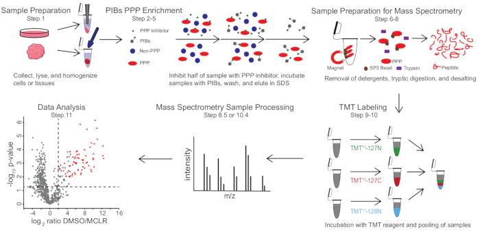
Figure 1: Visual summary of the PIB-MS protocol. In a PIB-MS experiment, samples can be obtained in various forms, from cells to tumors. The sample is collected, lysed, and homogenized prior to PPP enrichment. To enrich for PPPs, the lysate is incubated with PIBs with or without a PPP-inhibitor, such as MCLR. The PIBs are then washed, and PPPs are eluted in denaturing conditions. The samples are prepared for mass spectrometry analysis by the removal of detergents through SP3 protein enrichment, tryptic digestion, and desalting. Samples can then be optionally TMT-labeled prior to mass spectrometry analysis. Please click here to view a larger version of this figure.

PIB-MS involves lysis and clarification of cells or tissues, incubation of the lysate with PIBs, elution, and analysis of the eluate via western blotting or mass spectrometry-based approaches (Figure 1). The addition of free MCLR can be used as a control to distinguish specific PIB binders from non-specific interactors. For most applications, a label-free approach can be used to directly identify proteins in eluates. In cases where greater precision in quantification or the identification of low-abundance species is needed, further processing with tandem mass-tag (TMT) labeling can be used to increase coverage and decrease input.

Protocol

NOTE: The generation of PIBs is done as described by Moorhead et al., where 1 mg of microcystin and about 6 mL of sepharose are coupled to generate PIBs with a binding capacity of up to 5 mg/mL17. 1. Sample preparation NOTE: A typical starting amount for PIB-MS is 1 mg of total protein per condition. For this experiment, approximately 2.5 x 10⁶ HeLa cells were used to extract 1 mg of protein. This calculation should …

Representative Results

Figure 2: Identification of specific PIBs binders. (A) A variety of tissue types or cells can be analyzed via PIB-MS. HeLa cells in biological triplicate were either treated with DMSO or the PPP-inhibitor MCLR, incubated with PIBs, and analyzed via LC-MS/MS. (B) Volcano plot of PIB-MS anal…

Discussion

PIB-MS is a chemical proteomics approach used to quantitatively profile the PPPome from various sample sources in a single analysis. Much work has been done using kinase inhibitor beads to study the kinome and how it changes in cancer and other disease states10,11,12,13. Yet, the study of the PPPome lags behind. We anticipate that this approach is able to fill this gap and shed light on the reg…

Disclosures

The authors have nothing to disclose.

Acknowledgements

A.N.K. acknowledges support from NIH R33 CA225458 and R35 GM119455. We thank the Kettenbach and Gerber labs for their helpful discussion.

Materials

Acetonitrile (ACN) Honeywell AH015-4 CAUTION: ACN is flammable and toxic; wear gloves, and work in a chemical fume hood.
Anhydrous Acetonitrile Sigma-Aldrich 271004-100ML CAUTION: ACN is flammable and toxic; wear gloves, and work in a chemical fume hood.
Benchtop centrifuge Eppendorf model no. 5424
Beta-glycerophosphoric acid, disodium salt pentahydrate Acros Organics 410991000
Centrifuge Eppendorf model no. 5810 R 15 amp version
Distilled water
DMSO Fisher Scientific BP231-100
Dounce tissue grinder Fisherbrand Pellet Pestles 12-141-363
Empore solid phase extraction disk, C18 CDS Analytical 76333-132
Eppendorf tubes, 1.5 mL Eppendorf 22363204 CRITICAL: Other tubes may leach polymer into sample, contaminating the analysis.
Eppendorf tubes, 2 mL Eppendorf 22363352 CRITICAL: Other tubes may leach polymer into sample, contaminating the analysis.
Extraction plate manifold Waters WAT097944
Falcon tubes, 50 mL VWR 21008
Generic blunt end needle and plunger
Generic magnetic separation rack
HEPES Sigma-Aldrich H3375
Hydrogen chloride (HCl) VWR Chemicals BDH BDH3028 CAUTION: HCl is corrosive; wear gloves and work in a chemical fume hood.
Hydroxylamine solution 50% (wt/vol) Sigma-Aldrich 467804
Incubator, 65 °C VWR model no. 1380FM
Koptec Pure Ethanol, 200 Proof Decon Labs V1001
Methanol for HPLC (MeOH) Sigma-Aldrich 34860-4L-R CAUTION: MeOH is flammable and toxic; wear gloves, and work in a chemical fume hood.
Microcystin LR (MCLR) Cayman Chemical 10007188 CAUTION: MCLR is toxic; wear gloves when handling and avoid skin contact.
PBS, 1× without calcium and magnesium, pH 7.4 ± 0.1 Corning  21-040-CV
pH test strips, such as MilliporeSigma MColorpHast pH test strips and indicator papers Fisher Scientific M1095310001
PIBs For protocol for the generation of PIBs, see Moorhead et al., 2007.
Pierce BCA Protein Assay Kit Thermo Scientific 23225
Pipette tips, 10 μL Eppendorf 22491504 CRITICAL: Other tips may leach polymer into samples, contaminating the analysis.
Pipette tips, 1000 μL Eppendorf 22491555 CRITICAL: Other tips may leach polymer into samples, contaminating the analysis.
Pipette tips, 200 μL Eppendorf 22491539 CRITICAL: Other tips may leach polymer into samples, contaminating the analysis.
plastic syringe, 10 mL BD 309604
Protease inhibitor cocktail III Research Products International P50700-1
Q Exactive Plus Hybrid Quadrupole-Orbitrap Mass Spectrometer, Oribtrap Fusion, Orbitrap Fusion Lumos, or Orbitrap Eclipse Tribrid Mass Spectrometer  Thermo Scientific
Refrigerated benchtop centrifuge Eppendorf model no. 5424 R
Rotator (Labquake Shaker Rotisserie) Thermo Scientific 13-687-12Q 8 rpm rotation
Sample collection plate, 96- well, 1 mL Waters WAT058957
SDS Fisher Scientific BP1311-1
Sequencing grade modified trypsin Promega V511C
Sodium azide EMD Chemicals SX0299-1 CAUTION: Sodium azide is explosive and toxic; wear gloves, work in a chemical fume hood and avoid contact with metals.
Sodium chloride (NaCl) Fisher Chemical S27110
Sonicator (Branson digital sonifier) model no. SFX 250
SPE C18 desalting plate Waters 186001828BA
SpeedBeads magnetic carboxylate modified particles (SP3 beads) Cytiva 6.51521E+13
Thermomixer Eppendorf model no. 5350
TMT10plex Isobaric Label Reagent Set plus TMT11-131C Label Reagent, 3 × 0.8 mg per tag ThermoFisher A37725
Trifluoroacetic acid (TFA) Honeywell T6508-25ML CAUTION: TFA is corrosive and will irritate skin on contact. Wear gloves and eye protection, and work in a chemical fume hood.
Tris Base Research Products International T60040
Triton X-100 Sigma-Aldrich T9284
Vacuum centrifuge and vapor trap Thermo Scientific model nos. SpeedVac SPD120 and RVT5105
Vortexer (Vortex-Genie 2) Scientific Industries
Water LC-MS Honeywell LC365-4

References

  1. Nilsson, J. Protein phosphatases in the regulation of mitosis. Journal of Cell Biology. 218 (2), 395-409 (2019).
  2. Brautigan, D. L. Protein Ser/Thr phosphatases–the ugly ducklings of cell signalling. The FEBS Journal. 280 (2), 324-345 (2013).
  3. Brautigan, D. L., Shenolikar, S. Protein serine/threonine phosphatases: keys to unlocking regulators and substrates. Annual Review of Biochemistry. 87, 921-964 (2018).
  4. Janssens, V., Goris, J. Protein phosphatase 2A: A highly regulated family of serine/threonine phosphatases implicated in cell growth and signalling. Biochemical Journal. 353, 417-439 (2001).
  5. Virshup, D. M., Shenolikar, S. From promiscuity to precision: protein phosphatases get a makeover. Molecular Cell. 33 (5), 537-545 (2009).
  6. Bollen, M., Peti, W., Ragusa, M. J., Beullens, M. The extended PP1 toolkit: designed to create specificity. Trends in Biochemical Sciences. 35 (8), 450-458 (2010).
  7. Qian, J., Winkler, C., Bollen, M. 4D-networking by mitotic phosphatases. Current Opinion in Cell Biology. 25 (6), 697-703 (2013).
  8. Heroes, E., et al. The PP1 binding code: a molecular-lego strategy that governs specificity. The FEBS Journal. 280 (2), 584-595 (2013).
  9. Eichhorn, P. J., Creyghton, M. P., Bernards, R. Protein phosphatase 2A regulatory subunits and cancer. Biochimica et Biophysica Acta. 1795 (1), 1-15 (2009).
  10. Bantscheff, M., et al. Quantitative chemical proteomics reveals mechanisms of action of clinical ABL kinase inhibitors. Nature Biotechnology. 25 (9), 1035-1044 (2007).
  11. Klaeger, S., et al. Chemical proteomics reveals ferrochelatase as a common off-target of kinase inhibitors. ACS Chemical Biology. 11 (5), 1245-1254 (2016).
  12. Duncan, J. S., et al. Dynamic reprogramming of the kinome in response to targeted MEK inhibition in triple-negative breast cancer. Cell. 149 (2), 307-321 (2012).
  13. Cooper, M. J., et al. Application of multiplexed kinase inhibitor beads to study kinome adaptations in drug-resistant leukemia. PLoS ONE. 8 (6), 66755 (2013).
  14. Lyons, S. P., et al. A quantitative chemical proteomic strategy for profiling phosphoprotein phosphatases from yeast to humans. Molecular and Cellular Proteomics. 17 (12), 2448-2461 (2018).
  15. Nasa, I., et al. Quantitative kinase and phosphatase profiling reveal that CDK1 phosphorylates PP2Ac to promote mitotic entry. Science Signaling. 13 (648), (2020).
  16. Swingle, M., Ni, L., Honkanen, R. E. Small-molecule inhibitors of ser/thr protein phosphatases: specificity, use and common forms of abuse. Methods in Molecular Biology. 365, 23-38 (2007).
  17. Moorhead, G. B. G., Haystead, T. A. J., MacKintosh, C. Synthesis and use of the protein phosphatase affinity matrices microcystin-sepharose and microcystin-biotin-sepharose. Methods in Molecular Biology. 365, 39-45 (2007).
  18. Brauer, B. L., Wiredu, K., Mitchell, S., Moorhead, G. B., Gerber, S. A., Kettenbach, A. N. Affinity-based profiling of endogenous phosphoprotein phosphatases by mass spectrometry. Nature Protocols. 16 (10), 4919-4943 (2021).
  19. Hughes, C. S., Moggridge, S., Müller, T., Sorensen, P. H., Morin, G. B., Krijgsveld, J. Single-pot, solid-phase-enhanced sample preparation for proteomics experiments. Nature Protocols. 14 (1), 68-85 (2019).
  20. Rappsilber, J., Mann, M., Ishihama, Y. Protocol for micro-purification, enrichment, pre-fractionation and storage of peptides for proteomics using StageTips. Nature Protocols. 2 (8), 1896-1906 (2007).
  21. Zecha, J., et al. TMT labeling for the masses: A robust and cost-efficient, in-solution labeling approach. Molecular and Cellular Proteomics. 18 (7), 1468-1478 (2019).
  22. Elias, J. E., Gygi, S. P. Target-decoy search strategy for increased confidence in large-scale protein identifications by mass spectrometry. Nature Methods. 4 (3), 207-214 (2007).
  23. Eng, J. K., Jahan, T. A., Hoopmann, M. R. Comet: an open-source MS/MS sequence database search tool. Proteomics. 13 (1), 22-24 (2013).
  24. Tyanova, S., et al. The Perseus computational platform for comprehensive analysis of (prote)omics data. Nature Methods. 13 (9), 731-740 (2016).
  25. Yu, S. H., Ferretti, D., Schessner, J. P., Rudolph, J. D., Borner, G. H. H., Cox, J. Expanding the Perseus software for omics data analysis With custom plugins. Current Protocols in Bioinformatics. 71 (1), 1-29 (2020).
check_url/63805?article_type=t

Play Video

Cite This Article
Smolen, K. A., Kettenbach, A. N. A Mass Spectrometry-Based Approach to Identify Phosphoprotein Phosphatases and their Interactors. J. Vis. Exp. (182), e63805, doi:10.3791/63805 (2022).

View Video