We present a motor-powered centrifugal microfluidic device that can cultivate cell spheroids. Using this device, spheroids of single or multiple cell types could be easily cocultured under high gravity conditions.
A three-dimensional spheroid cell culture can obtain more useful results in cell experiments because it can better simulate cell microenvironments of the living body than two-dimensional cell culture. In this study, we fabricated an electrical motor-driven lab-on-a-CD (compact disc) platform, called a centrifugal microfluidic-based spheroid (CMS) culture system, to create three-dimensional (3D) cell spheroids implementing high centrifugal force. This device can vary rotation speeds to generate gravity conditions from 1 x g to 521 x g. The CMS system is 6 cm in diameter, has one hundred 400 μm microwells, and is made by molding with polydimethylsiloxane in a polycarbonate mold premade by a computer numerical control machine. A barrier wall at the channel entrance of the CMS system uses centrifugal force to spread cells evenly inside the chip. At the end of the channel, there is a slide region that allows the cells to enter the microwells. As a demonstration, spheroids were generated by monoculture and coculture of human adipose-derived stem cells and human lung fibroblasts under high gravity conditions using the system. The CMS system used a simple operation scheme to produce coculture spheroids of various structures of concentric, Janus, and sandwich. The CMS system will be useful in cell biology and tissue engineering studies that require spheroids and organoid culture of single or multiple cell types.
It is easier to simulate biological in vivo microenvironments with three-dimensional (3D) spheroid cell culture than with two-dimensional (2D) cell culture (e.g., conventional Petri dish cell culture) to produce more physiologically realistic experimental results1. Currently available spheroid formation methods include the hanging drop technique2, liquid-overlay technique3, carboxymethyl cellulose technique4, magnetic force-based microfluidic technique5, and the use of bioreactors6. Although each method has its own benefits, further improvement in reproducibility, productivity, and generating coculture spheroids is necessary. For example, while the magnetic force-based microfluidic technique5 is relatively inexpensive, the effects of strong magnetic fields on living cells must be carefully considered. The benefits of spheroid culture, particularly in the study of mesenchymal stem cell differentiation and proliferation, have been reported in several studies7,8,9.
The centrifugal microfluidic system, also known as lab-on-a-CD (compact disc), is useful for easily controlling the fluid inside and exploiting the rotation of the substrate and has thus been utilized in biomedical applications such as immunoassays10, colorimetric assays for detecting biochemical markers11, nucleic acid amplification (PCR) assays, automated blood analysis systems12, and all-in-one centrifugal microfluidic devices13. The driving force controlling the fluid is the centripetal force created by rotation. Additionally, multiple functions of mixing, valving, and sample splitting can be done simply in this single CD platform. However, compared to the above-mentioned biochemical analysis methods, there have been fewer trials applying CD platforms to culture cells, especially spheroids14.
In this study, we show the performance of the centrifugal microfluidic-based spheroid (CMS) system by monoculture or coculture of human adipose-derived stem cells (hASC) and human lung fibroblasts (MRC-5). This paper describes in detail our group's research methodology15. Thus, the spheroid culture lab-on-a-CD platform can be easily reproduced. A CMS generating system comprising a CMS culture chip, a chip holder, a DC motor, a motor mount, and a rotating platform, is presented. The motor mount is 3D printed with acrylonitrile butadiene styrene (ABS). The chip holder and rotating platform are CNC (computer numerical control) machined with the PC (polycarbonate). The rotational speed of the motor is controlled from 200 to 4,500 rpm by encoding a PID (proportional-integral-derivative) algorithm based on pulse-width modulation. Its dimensions are 100 mm x 100 mm x 150 mm and it weighs 860 g, making it easy to handle. Using the CMS system, spheroids can be generated under various gravity conditions from 1 x g to 521 x g, so the study of cell differentiation promotion under high gravity can be extended from 2D cells16,17 to 3D spheroid. Coculture of various types of cells is also a key technology for effectively mimicking the in vivo environment18. The CMS system can easily generate monoculture spheroids, as well as coculture spheroids of various structure types (e.g., concentric, Janus, and sandwich). The CMS system can be utilized not only in simple spheroid studies but also in 3D organoid studies, to consider human organ structures.
1. Centrifugal microfluidic-based spheroid (CMS) culture chip fabrication
2. Cell preparation
3. Monoculture spheroid formation
4. Coculture spheroid formation
5. Cell staining
The 6 cm diameter CMS culture chip (Figure 2) was successfully made following the above protocol. First, the chip was made separately from a top layer and a bottom layer and then bonded together by plasma bonding. Resulting spheroids can be easily gathered by detaching the chip. The channel of the CMS culture chip comprises an inlet port and central, slide, and microwell regions (Figure 3). The cell, medium, and pluronic solutions are injected through an inlet hole with a diameter of 5 mm. The injected cells are evenly distributed by resuspension 3–5x in the inlet port region. The cells are subjected to centrifugal force in the central region and spread outward. Because the central region is higher than the microwell, it can contain more media, allowing the spheroids to survive longer. The height of the microwell is 0.4 mm and the height of the central region is 1.5 mm. Suction holes are present at the center of the central region to easily remove the internal solutions. The slide region is a sloping area connecting the central region and the microwell region. The cells move along a 45° slope and settle in the microwell region, where the cells settle, grow, and tangle to form spheroids. Microwells located 14 mm from the center of the chip are semicylindrical with a height of 400 μm and a diameter of 400 μm. A total of 100 spheroids can be generated simultaneously in 100 microwells.
Using the prepared CMS chip, spheroids can be generated in the order shown in the protocol (Figure 4). Monoculture and coculture spheroids were generated with hASC and MRC-5 cells. To generate a monoculture spheroid of each type of cell, 5 × 10⁵ hASCs or 8 × 10⁵ MRC-5s were injected. The number of cells injected was independent of the cell size. Time-lapse images of both types of cells were taken at 2,000 rpm until day 3 of cell culture (Figure 5). Coculture spheroids of hASCs and MRC-5s were also generated with concentric, Janus, and sandwich structures. To make concentric spheroids, the first cells (2.5 × 10⁵ hASCs) were injected and the second cells (4 × 10⁵ MRC-5s) were injected 3 min later (Figure 6A). When the first cells were injected, they became U-shaped due to high gravity, and when the second cells were injected, they moved to the middle of the U-shape. Over time, the first U-shape cells encircled the second cells and completed the concentric spheroids. To make Janus spheroids, the first cells (2.5 × 10⁵ hASCs) were injected, and the second cells (4 × 10⁵ MRC-5s) were injected 3 h later (Figure 6B). When the injection interval between the two cells was long, the shape of the first cells changed from a U-shape to an elliptical shape by cell aggregation. Once the second cells were added to the elliptical shape of the first cells, the Janus spheroids were generated. In the case of the sandwich spheroids, the first cells (1.5 × 10⁵ hASCs) were injected, the second cells (3 × 10⁵ MRC-5s) were injected 3 h later, and the third cells (1.5 × 10⁵ hASCs) were injected after another 3 h (Figure 6C). Similar to the Janus spheroid, each cell aggregated into an elliptical shape, and the three layers stacked to generate sandwich spheroids. Lastly, to demonstrate the long-term culture capability of the CMS, hASCs were cultured, exposed to high gravity for 7 days followed by a live/dead assay performed to show that most cells survived (Figure 7). Also, photos of all microwells of the CMS were taken after 3 days of MRC-5s cultivation to show excellent uniformity and sphericity of spheroids (Figure 8).
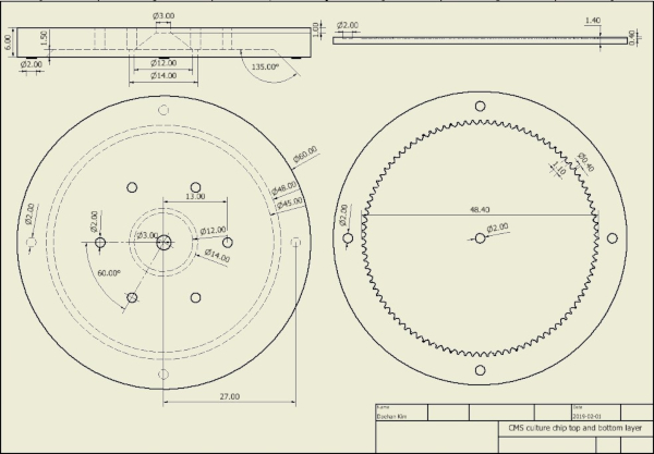
Figure 1: Dimensions of the top and bottom layers of a CMS culture chip. The PC mold was made using a CNC machine and replicated with PDMS to make a CMS culture chip based on the drawing created by a 3D CAD (computer-aided design) program. The four circles at the edges of the top and bottom layers are for aligning the two layers. Dimensions are in millimeters. Please click here to view a larger version of this figure.
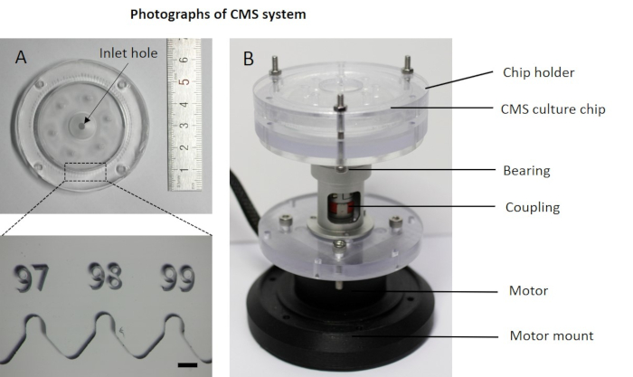
Figure 2: Photographs of the CMS system. (A) Photographs of the completed CMS culture chip. The diameter of the chip is 6 cm and the diameter of the microwell is 400 μm. The numbers above the microwells represent the individual numbers of the microwells from 1 to 100. These numbers were engraved into the mold. Scale bar = 400 μm. (B) Photograph of the whole CMS system. The CMS system comprises the CMS culture chip, chip holder, DC motor, and rotating platform. CMS devices can generate gravity conditions up to 521 x g through rotational force. The chip holder prevents separation of the CMS culture chip due to high gravity. Please click here to view a larger version of this figure.
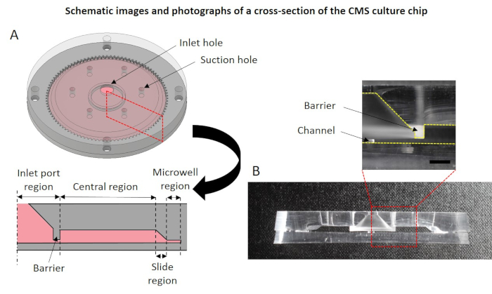
Figure 3: Channel components of the CMS. (A) Schematic images and (B) photographs of a cross-section of the CMS culture chip. The CMS culture chip consists of an inlet port and central, slide, and microwell regions. Because the injected cells do not pass through the barrier at a rotational speed of less than 1,000 rpm, the barrier helps in the resuspension of the cell and even distribution to the microwell. Scale bar = 2 mm. Please click here to view a larger version of this figure.
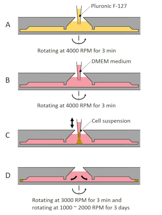
Figure 4: Process of loading cells into the CMS culture chip. (A) To prevent cells from sticking to the bottom of the chip, coat with 2.5 mL of the pluronic F-127 solution at 4,000 rpm. Wait a day for the coating to be applied. (B) Remove the pluronic solution and prefill the channel with 2.5 mL of the DMEM medium. (C) Remove 100 μL of the DMEM and add 100 μL of cell suspension. At this time, resuspend 3–5x so that the cells are evenly distributed. (D) Move the cells to the microwell by rotating the chip, and then culture the cells for 3 days at 1,000 to 2,000 rpm. Please click here to view a larger version of this figure.
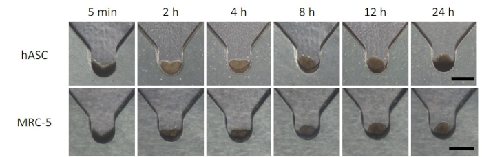
Figure 5: Time-lapse photograph of monoculture spheroids of hASC and MRC-5 cells. Cells were grown for 24 h at 2,000 rpm. The spheroid was generated within 24 h. Scale bar = 400 μm. Please click here to view a larger version of this figure.
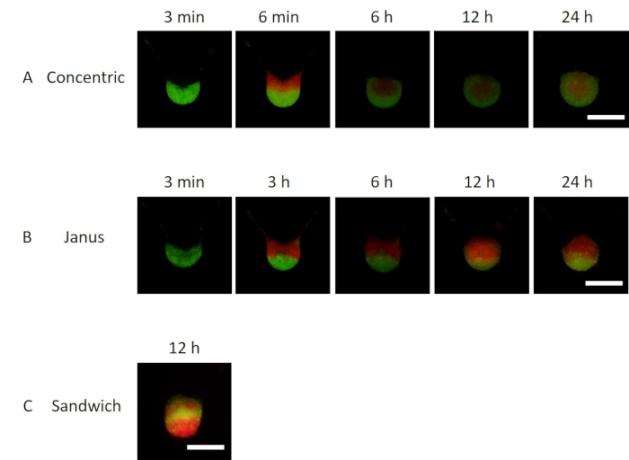
Figure 6: Fluorescence images of coculture spheroids. (A) Concentric spheroid shapes in which hASC cells (green) surround MRC-5 cells (red). (B) Janus spheroid shape in which two cells are symmetrical. (C) Sandwich spheroid shape in which hASC layers are stacked between two MRC-5 layers. Scale bar = 400 μm. Please click here to view a larger version of this figure.
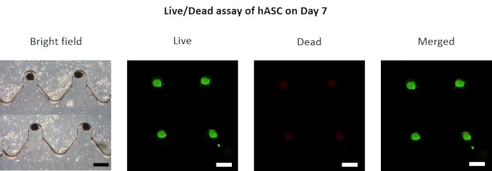
Figure 7: Live/Dead assay of hASC on Day 7. The green fluorescent color represents living cells and the red fluorescent color represents dead cells. Scale bar = 400 μm. Please click here to view a larger version of this figure.
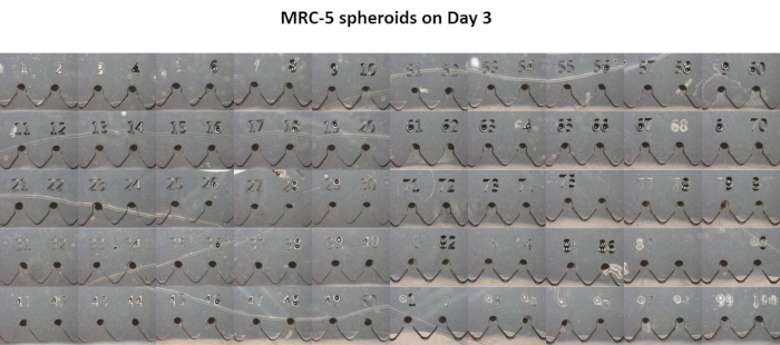
Figure 8: MRC-5 spheroids on Day 3. A relatively constant number of cells enter each microwell and form spheroids having relatively constant sphericity in the CMS system. Please click here to view a larger version of this figure.
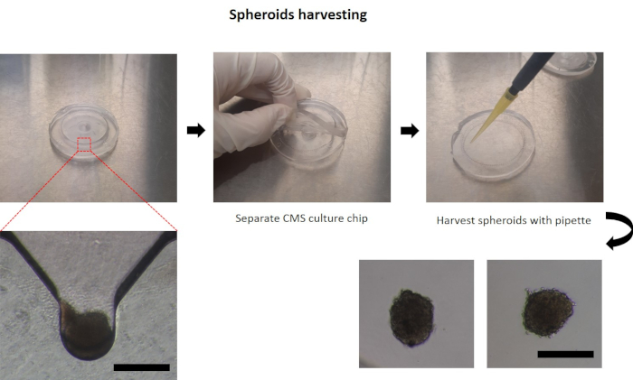
Figure 9: Harvesting spheroids. Cultured spheroids can be harvested by dividing the two layers of the CMS culture chip. The two plasma-bonded layers can be easily separated by hand. Then the spheroids are collected from the microwells in the bottom layer simply by pipetting. Scale bar = 400 μm. Please click here to view a larger version of this figure.
The CMS is a closed system in which all injected cells enter the microwell without waste, making it more efficient and economical than conventional microwell-based spheroid generation methods. In the CMS system, the media is replaced every 12–24 h through a suction hole designed to remove the media in the chip (Figure 3A). During the media suction process, barely any media escapes from inside the microwell due to the surface tension between the media and the wall of the microwell. A user can easily remove the trapped media by pressing near the microwell region of the chip with a finger, because the chip made of PDMS is elastic and flexible. Cells in the microwell remain stable without escaping, even with multiple media changes. To achieve the same quality of spheroid in all 100 microwells, the rotation of the device should not be eccentric, and the chip should be axisymmetrical. Otherwise, a variable number of cells may enter each well and the size and shape of the spheroid could differ. In the conventional microwell system, because the air is often trapped in the microwell, it is necessary to remove the air bubbles. However, the CMS system does not require the bubble removal process because the high centrifugal force generated by the rotation causes the media to push the bubbles and squeeze them out from the microwells.
The CMS system also has its limitations compared to conventional methods. The CMS system requires a larger culture space (e.g., large incubator space) as it comprises a motor, rotating platform, and a controller, and its total size is approximately 100 mm x 100 mm x 150 mm (Figure 2B). In addition, it causes a slight but persistent vibration. We expect that the miniaturization of the system (hopefully similar to the size of 6 well plates) will solve these issues.
It should be noted that the cultured spheroids can be collected by separating the two plasma-bonded layers of the CMS system (Figure 9). The bonding of the two layers is strong enough to prevent the media from leaking during system operation. However, owing to the small bonding area, it is separable by hand. The spheroids can be simply collected from the bottom layer by pipetting.
The CMS system has better reproducibility and productivity than conventional methods of spheroid generation. The conventional methods, such as normal microwell or hanging droplet methods, are labor-intensive. However, in the CMS system, it is much easier to increase the number of spheroids by simply increasing the chip size. With this device, it is also possible to generate organoids that require culture of multicell types, which is not easy to do in conventional culture methods. In addition to the cells in this study (hASC and MRC-5), CMS could be used for spheroid production using various other types of cells that can form spheroids.
The authors have nothing to disclose.
This research was supported by the Basic Science Research Program (2016R1D1A1B03934418) and the Bio & Medical Technology Development Program (2018M3A9H1023141) of the NRF, and funded by the Korean government, MSIT.
3D printer | Cubicon | 3DP-210F | |
Adipose-derived mesenchymal stem cells (hASC) | ATCC | PCS-500-011 | |
Antibiotic-Antimycotic | Gibco | 15240-062 | Contained 1% of completed medium and buffer |
CellTracker Green CMFDA | Thermo Fisher Scientific | C2925 | 10 mM |
CellTracker Red CMTPX | Thermo Fisher Scientific | C34552 | 10 mM |
Computer numerical control (CNC) rotary engraver | Roland DGA | EGX-350 | |
DC motor | Nurielectricity Inc. | MB-4385E | |
Dimethylsulfoxide (DMSO) | Sigma Aldrich | D2650 | |
Dulbecco's modified eaggle's medium (DMEM) | ATCC | 30-2002 | |
Dulbecco's phosphate buffered saline (D-PBS) | ATCC | 30-2200 | |
Fetal bovine serum | ATCC | 30-2020 | Contained 10% of completed medium |
human lung fibroblasts (MRC-5) | ATCC | CCL-171 | |
Inventor 2019 | Autodesk | 3D computer-aided design program | |
Petri dish Φ 150 mm | JetBiofill | CAD010150 | Surface Treated |
Plasma cleaner | Harrick Plasma | PDC-32G | |
Pluronic F-127 | Sigma Aldrich | 11/6/9003 | Dilute with phosphate buffered saline to 4% (w/v) solution |
Polycarbonate (PC) | Acrylmall | AC15PC | 200 x 200 x 15 mm |
Polydimethylsiloxane (PDMS) | Dowcorning | Sylgard 184 | |
Trypsin | Gibco | 12604021 |