JoVE Science Education
Microbiology
A subscription to JoVE is required to view this content.  Sign in or start your free trial.
JoVE Science Education Microbiology
Plaque Assay: A Method to Determine Viral Titer as Plaque Forming Units (PFU)
  • 00:01Concepts
  • 03:09Media Preparation
  • 05:12Preparing Agar Plates
  • 06:21Culturing Host Cells
  • 07:30Phage Serial Dilution and Preparation of Bacteria and Phage Overlay
  • 10:37Data Analysis and Results
  • 11:35Results

Test de la plaque : méthode de détermination de la charge virale exprimée en unités formant des plaques

English

Share

Overview

Source: Tilde Andersson1, Rolf Lood1
1 Département des sciences cliniques Lund, Division of Infection Medicine, Biomedical Center, Lund University, 221 00 Lund, Suède

Les virus qui infectent les organismes procaryotes, appelés bactériophages ou simplement phages, ont été identifiés au début duXXe siècle par Twort (1) et d’Hérelle (2) indépendamment. Les phages ont depuis été largement reconnus pour leur valeur thérapeutique (3) et leur influence sur les écosystèmes humains (4), ainsi que sur les écosystèmes mondiaux (5). Les préoccupations actuelles ont alimenté un regain d’intérêt pour l’utilisation des phages comme alternative aux antibiotiques modernes dans le traitement des maladies infectieuses (6). Essentiellement, toutes les recherches sur les phages reposent sur la capacité de purifier et de quantifier les virus, également connu sous le nom de titre viral. Initialement décrit en 1952, c’était le but de l’assiduité de la plaque (7). Des décennies et de multiples progrès technologiques plus tard, l’analyse de la plaque demeure l’une des méthodes les plus fiables pour la détermination du titre viral (8).

Les bactériophages subsisent en injectant leur matériel génétique dans les cellules hôtes, en détournant les machines pour la production de nouvelles particules de phage, et finalement en provoquant l’hôte de libérer de nombreuses virions progéniture par lyse cellulaire. En raison de leur taille infime, les bactériophages ne peuvent pas être observés à l’aide uniquement de microscopie légère; par conséquent, la microscopie électronique de balayage est nécessaire (figure 1). En outre, les phages ne peuvent pas être cultivés sur les plaques d’agar nutritionnelles comme les bactéries, car ils ont besoin de cellules hôtes pour s’en prendre à eux.

Figure 1
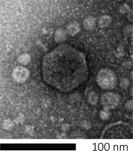
Figure 1 : La morphologie d’un bactériophage, ici illustrée par un phage E. coli, peut être étudiée à l’aide de la microscopie électronique à balayage. La plupart des bactériophages appartiennent aux Caudovirales (bactériophages à queue). Ce phage particulier a une structure de queue très courte et une tête icosaèdre, le plaçant dans la famille des Podovirus.

L’essai de plaque (figure 2) est basé sur l’incorporation des cellules hôtes, préférentiellement dans la croissance de phase de journal, dans le milieu. Cela crée une couche dense et turbide de bactéries capables de soutenir la croissance virale. Un phage isolé peut ensuite infecter, se répliquer et lyser une cellule. Avec chaque cellule lysée, plusieurs cellules adjacentes deviennent immédiatement infectées. Plusieurs cycles dans, une zone claire (une plaque) peut être observée dans la plaque autrement turbide (figure 2B/figure 3A), indiquant la présence de ce qui était initialement une particule bactériophage unique. Le nombre d’unités de formation de plaques par volume(c.-à-d. PFU/mL) d’un échantillon, peut donc être déterminé à partir du nombre de plaques générées.

Figure 2
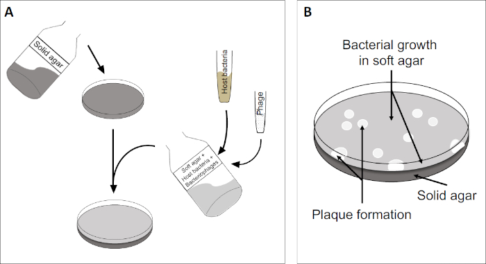
Figure 2 : Le dépistage des unités de formation de plaque (PFU) est une méthode courante pour déterminer le nombre de bactériophages dans un échantillon. (A) La base d’un plat stérile Petri est recouverte d’un milieu nutritif solide approprié, suivie d’un mélange de milieux mous, de cellules hôtes sensibles et d’une dilution de l’échantillon de bactériophage d’origine. Notez que la suspension de phage pourrait, dans certains cas, également être répartie uniformément sur la surface de l’agar mou déjà solidifié. (B) La croissance des bactéries hôtes forme une pelouse de cellules dans la couche supérieure d’agar. La réplication des bactériophages génère des zones claires, ou plaques, causées par la lyse des cellules hôtes.

Figure 3
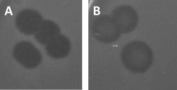
Figure 3 : Les résultats des tests PFU montrent plusieurs plaques générées par des bactériophages. En raison de la lyse des cellules hôtes sensibles, les plaques peuvent être considérées comme des zones de dégagement dans la pelouse bactérienne, soit avec (A) dégagement complet, soit (B) re-croissance partielle causée par la génération de bactéries résistantes (ou peut-être par des phages tempérés dans le cycle lysogénique).

Certains phages tempérés peuvent adopter ce qu’on appelle un cycle de vie lysogénique, en plus de la croissance lytique précédemment décrite. En lysogénie, le virus assume un état latent par l’incorporation de son matériel génétique dans le génome de la cellule hôte (9), conférant souvent une résistance à d’autres infections phages. Cela se révèle parfois par un léger obscurcissement de la plaque (figure 3B). Il est intéressant de noter cependant, que les plaques peuvent également apparaître floues en raison de la repousse des bactéries qui ont évolué résistance au phage indépendamment des infections phage précédente.

Les virus peuvent se fixer, ou adsorb, à seulement une gamme limitée de bactéries hôtes (10). Les plages d’hôtes sont encore limitées par des stratégies antivirales intracellulaires telles que le système CRISPR-Cas (11). La résistance/sensibilité à l’égard de phages spécifiques affichés par les sous-groupes bactériens a toujours été utilisée pour classer les souches bactériennes en différents types de phages (figure 4). Bien que l’efficacité de cette méthode ait maintenant été surpassée par de nouvelles techniques de séquençage, la dactylographie peut encore fournir des informations précieuses sur les interactions bactéries-phage, par exemple, en facilitant la conception d’un cocktail de phage pour une utilisation clinique .

Figure 4
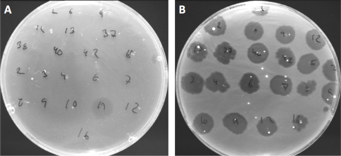
Figure 4 : Sensibilité aux phages de différentes souches bactériennes. Des plaques d’agar molles avec la souche d’acné de Cutibacterium (A) AD27 et (B) AD35, ont été repérées avec 21 bactériophages différents de C. acnes. Seul phage 11 a été en mesure d’infecter et de tuer AD27 tandis que la souche AD35 a montré une sensibilité envers tous les phages. Cette technique, appelée dactylographie phage, peut être utilisée pour diviser les espèces bactériennes et les souches en différents sous-groupes basés sur la susceptibilité au phage.

Procedure

1. Mise en place Avant de commencer tout travail impliquant des microbes, assurez-vous que l’espace de travail est stérilisé(p. ex. essuyé avec 70 % d’éthanol). Portez toujours une blouse de laboratoire et des gants, gardez les cheveux longs attachés en arrière et assurez-vous que les plaies sont particulièrement bien protégées. Une fois terminé, stériliser toutes les surfaces et bien laver /stériliser les mains et les poignets. <st…

Applications and Summary

Despite multiple technological advances, plaque assays remain the gold standard for determination of viral titer (as PFU) and essential for isolation of pure bacteriophage populations. Susceptible host cells are cultivated in the top coat of a two layered agar-plate, forming a homogenous bed enabling viral replication. The initial event where an isolated bacteriophage in lytic lifecycle infects a cell, replicates within it, and eventually lyses it, is too small to observe. However, the virions released will infect adjacent cells, subsequently giving rise to a clearing zone, or plaque, denoting the presence of a single PFU.

References

  1. Twort, F. An investigation on the nature of ultra-microscopic viruses. Lancet. 186 (4814): 1241-1243. (1915)
  2. d'Hérelle, F. An invisible antagonist microbe of dysentery bacillus. Comptes Rendus Hebdomadaires Des Seances De L Academie Des Sciences. 165: 373-375. (1917)
  3. Cisek AA, Dąbrowska I, Gregorczyk KP, Wyżewski Z. Phage Therapy in Bacterial Infections Treatment: One Hundred Years After the Discovery of Bacteriophages. Current Microbiology. 74 (2):277-283. (2017)
  4. Mirzaei MK, Maurice CF. Ménage à trois in the human gut: interactions between host, bacteria and phages. Nature Reviews Microbiology. 15 (7):397. (2017)
  5. Breitbart M, Bonnain C, Malki K, Sawaya NA. Phage puppet masters of the marine microbial realm. Nature Microbiology. 3 (7):754-766. (2018)
  6. Leung CY, Weitz JS. Modeling the synergistic elimination of bacteria by phage and the innate immune system. Journal of Theoretical Biology. 429:241-252. (2017)
  7. Dulbecco R. Production of Plaques in Monolayer Tissue Cultures by Single Particles of an Animal Virus. Proceedings of the National Academy of Sciences of the United States of America. 38 (8):747-752. (1952)
  8. Juarez D, Long KC, Aguilar P, Kochel TJ, Halsey ES. Assessment of plaque assay methods for alphaviruses. J Virol Methods. 187 (1):185-9. (2013)
  9. Clokie MRJ, Millard AD, Letarov AV, Heaphy S. 2011. Phages in nature. Bacteriophage. 1 (1):31-45. (2011)
  10. Moldovan R, Chapman-McQuiston E, Wu XL. On kinetics of phage adsorption. Biophys J. 93 (1):303-15. (2007)
  11. Garneau JE, Dupuis M-È, Villion M, Romero DA, Barrangou R, Boyaval P, Fremaux C, Horvath P, Magadán AH, Moineau S.. The CRISPR/Cas bacterial immune system cleaves bacteriophage and plasmid DNA. Nature. 468 (7320):67. (2010)

Transcript

Bacteriophages, also called phages, are viruses that specifically infect bacteria and we can confirm their presence and quantify them using a tool called the plaque assay. Bacteriophages infect their susceptible hosts by first attaching to the bacterial cell wall and injecting their genetic material. Then, they hijack the cell’s biosynthetic machinery to replicate their DNA and produce numerous progeny phage particles, which they then release by lysing and killing the host cell.

This lytic activity is the basis of a widely used phage enumerating technique known as the plaque assay or double agar layer assay. Here, a bacteriophage mix is first prepared in a molten nutrient broth containing low concentration agar. All bacteria used in the mix should be alive and actively dividing in the log phase of their growth, which will ensure that a large percentage of the bacteria are viable and able to form a dense lawn around the plaques. Next, this molten bacterial-phage agar mix is spread over a more solid, concentrated agar nutrient medium which is already solidified on a Petri dish. On incubation at room temperature, the low concentration agar-phage-bacteria broth also solidifies to form a soft agar overlay.

Here, the bacterial cells can derive additional nutrients from the bottom layer and should rapidly multiply to produce a confluent lawn of bacteria. However, as phage particles are also present in the soft layer, these will infect and replicate their genetic material within the bacteria, culminating in cell lysis, which releases multiple progeny. These phage progeny then infect the neighboring cells, as the semi-solid state of the bacteria-phage layer restricts their movement to more distantly located host cells. This cycle of infection and lysis continues over multiple rounds, killing a large number of bacteria in a localized area. The effect of the neighboring cells being destroyed, is to produce a single circular clear zone, called a plaque, which can be seen by the naked eye, effectively amplifying the bacteria lytic activity of the phage and enabling their enumeration.

The number of plaques on a Petri dish are referred to as Plaque-Forming Units, or PFUs, and, providing the initial bacteriophage concentration was sufficiently dilute, should directly correspond to the number of infective phage particles in the original sample. This technique can also be used for characterization of plaque morphology, to aid in identification of phage types, or to isolate phage mutants. In this lab, you will learn how to perform the plaque assay for enumerating phages, using the T7 phage of E. coli as an example.

First, identify a suitable medium for the culturing of the host bacterial cells and the bacteriophage. Here lysogeny broth, or LB medium, was used to culture E. coli and the T7 phage. Next, take three clean glass bottles and label them with media, name, and then the first as LB-Broth, the second as LB-Bottom Agar, and the third as LB-Top Agar. Now, weigh out four grams of pre-formulated LB powder in three sets and then transfer one set of weighed dried media into each bottle. Add 200 milliliters of water to the first bottle. Mix the contents using a magnetic stir bar.

Then, using a pH meter and constant stirring, bring the final pH to 7.4 through the addition of sodium hydroxide or hydrochloric acid. Repeat the water addition and pH adjustment for the other two remaining bottles, as well. Now, weigh out three grams of agar powder and add it to the second bottle to make a 1.5 % bottom agar. Finally, weigh 1. 2 grams of agar and add it to the third bottle to make the .6 % LB top agar. The broth condition in bottle one does not need an agar addition. Cap the bottle semi-tightly and then, sterilize the media by autoclaving at 121 degrees Celsius for 20 minutes. Once complete, remove the media bottles from the autoclave and immediately twist the bottle caps to close them fully to prevent contamination. Keep the LB-Broth and LB-Top Agar media on the bench for later use. Place the LB-Bottom Agar to cool in a water bath that is preset to approximately 45 degrees Celsius.

When the LB-Bottom agar reaches approximately 45 degrees Celsius, transfer it to the work bench. Next, sterilize the workspace using 70 % ethenol. Next, add 450 microliters of sterile one molar calcium chloride to the molten bottom agar to make a final concentration of 2.25 millimolar. Gently swirl the bottle to mix. Then, set out seven clean Petri dishes. Label each dish on the bottom with the media name and preparation date. Then, pour 15 milliliters of the bottom agar into each of the seven Petri dishes. Allow the plates to set for a few hours or overnight at room temperature. Once set, the culture plates can be stored at four degrees Celsius for several days if needed, upside down to minimize condensation. Transfer the Petri dishes from the four degrees Celsius refrigerator to a 37 degrees Celsius incubator one hour before the assay.

The day before the assay is to be preformed, the E. coli should be cultured. Here, 10 microliters of E. coli culture was inoculated into 10 milliliters of LB-Broth. Place the bacteria to grow overnight in a shaking incubator set to 37 degrees Celsius at 160 RPM. Then, on the day of the assay, remove the bacterial culture from the incubator. Seed a fresh 10 milliliters of fresh LB broth with 0.5 milliliters of the overnight culture. Place these cells to grow into a shaking incubator set to 37 degrees Celsius at 160 RPM. Next, use a spectrophotometer to check when this culture reaches log phase growth, indicated by an optical density of 0.5 to 0.7. Once the OD reaches this level, stop the incubation by transferring the cell culture to the bench. They are now ready to be used for phage overlay assay.

Phage titers can vary exponentially across different phage types and samples. So in order to count them effectively, they should be diluted to generate a wide range of phage concentrations. On the day of the assay, generate a series of phage dilutions ranging from one tenth to one millionth concentrations, following a 10-fold dilution technique. To obtain statistically significant and accurate data, perform the serial dilution in triplicate.

Next, melt the solidified LB-top agar using a microwave. Then, place it in a water bath that is preset at 45 degrees Celsius for one hour. After one hour, collect the Petri dishes containing the bottom agar layer from the incubator. Label the plates with phage concentration and assay date. Then, set out seven clean test tubes. Label each test tube with the serial phage dilution number and designate one as control.

When the LB-top agar reaches 45 degrees Celsius, transfer it to the working bench. Now, add 450 microliters of one molar calcium chloride to the 200 milliliter agar to make a final concentration of 2.25 millimolar. Gently swirl the bottle to mix. Next, add 35 milliliters of LB-top agar and four milliliters of bacterial suspension to a sterile conical tube. Gently swirl to evenly distribute the cells but avoid shaking to prevent foaming.

Now, aliquot five milliliters of this bacteria- top agar mix to each of the seven test tubes. Then, transfer 100 microliters of the serially diluted bacteriophage samples and control media, which should be simply media with no bacteriophage, to the respectfully labeled test tubes. Swirl the mixture gently to ensure proper mixing. Gently transfer five milliliters of bacteriophage mix onto the respective Petri plate. Evenly spread the mix throughout the whole surface by gently swirling the Petri plate.

Once all of the Petri plates are layered with the mix, allow solidification of the top layer by incubating at room temperature for 15 minutes. After completion of these steps, repeat the process for the second and then the third sets of the Petri dishes using the remaining two sets of phage dilutions. Seal each dish with parafilm and incubate at room temperature for 15 minutes. Place the culture plate upside down at a suitable temperature for 24 hours or until plaques develop. Here, plates were placed in a 37 degrees Celsius incubator for one day, a stimulating growth condition for E. coli and the T7 phage.

Plaques will appear after one to five days of incubation, depending on the bacterial species, incubation conditions, and the choice of medium. Here, plaques were visible after one day of incubation at 37 degrees Celsius. Begin by checking the plates marked control and ensure that no plaques were formed in these plates, as this would indicate viral contamination. To determine the phage titer in the original sample, start with the plates containing the most diluted phage sample first and count the plaques without removing the lids, marking them to indicate which ones have already been counted. Repeat the counting for each plate in every set. Some plates might have too many or too few plaques to be counted. Consider 10 to 150 as an ideal plaque count.

Next, generate a table listing the plaque number values for the different dilutions and replicates. Then, calculate the mean plaque number values for the dilution plates that contained the ideal number of plaque counts. In this example, these were the average number of plaques formed in the 10 to the minus three and 10 to the minus four dilution plates. Now, adjust for phage dilution factor by dividing the obtained mean plaque values by the respective phage dilution factors. Here, the average number of plaques formed to the 10 to the minus three and 10 to the minus four dilution plates, were divided by their respective dilution factors to obtain the number of plaque forming units, or PFUs, in 100 microliters of phage mixture. To convert the value to PFU per milliliter, multiply the generated values by 10, as only 100 microliters of phage dilution mix was used during the bacteriophage overlay preparation step, producing an additional dilution factor of 10. Finally, calculate the average of the values obtained from the different dilution plates. This will give the average number of PFUs per milliliter. The number of PFUs corresponds to the number of infective phage particles in the original sample.

Tags

Cite This
JoVE Science Education Database. JoVE Science Education. Plaque Assay: A Method to Determine Viral Titer as Plaque Forming Units (PFU). JoVE, Cambridge, MA, (2023).