The present protocol describes a method for injecting tick embryos. Embryo injection is the preferred technique for genetic manipulation to generate transgenic lines.
Ticks can transmit various viral, bacterial, and protozoan pathogens and are therefore considered vectors of medical and veterinary importance. Despite the growing burden of tick-borne diseases, research on ticks has lagged behind insect disease vectors due to challenges in applying genetic transformation tools for functional studies to the unique biology of ticks. Genetic interventions have been gaining attention to reduce mosquito-borne diseases. However, the development of such interventions requires stable germline transformation by injecting embryos. Such an embryo injection technique is lacking for chelicerates, including ticks. Several factors, such as an external thick wax layer on tick embryos, hard chorion, and high intra-oval pressure, are some obstacles that previously prevented embryo injection protocol development in ticks. The present work has overcome these obstacles, and an embryo injection technique for the black-legged tick, Ixodes scapularis, is described here. This technique can be used to deliver components, such as CRISPR/Cas9, for stable germline transformations.
Ticks are vectors of medical and veterinary importance, capable of transmitting a variety of viral, bacterial, protozoan pathogens and nematodes1,2. In the eastern United States, the black-legged tick, Ixodes scapularis, is an important vector of the Lyme disease (LD) pathogen, the spirochete Borrelia burgdorferi. Over 400,000 cases of LD are reported each year in the United States, making it the top vector-borne infectious disease in the US1. In addition to B. burgdorferi, six other microorganisms are transmitted by I. scapularis– including four bacteria (Anaplasma phagocytophilum, B. mayonii, B. miyamotoi, and Ehrlichia muris eauclarensis), one protozoan parasite (Babesia microti), and one virus (Powassan virus), making this tick species a major public health concern3. While tick-borne diseases have become more prevalent in recent years, research on ticks has fallen behind other arthropod vectors, such as mosquitoes, due to the unique biology of ticks and challenges associated with applying genetic and functional genomic tools4,5.
Gene-editing techniques, particularly CRISPR/Cas9, have now made functional genomics studies feasible in non-model organisms. For creating heritable mutations in an organism, embryo injection remains the preferred method for delivering constructs for altering the germline6,7,8,9. However, until recently4, tick eggs were considered too difficult or even impossible to inject without killing the embryo10,11. A thick wax layer on eggs, hard chorion, and high intra-oval pressure were some of the main obstacles that prevented embryo injection in ticks. Adult, blood-fed I. scapularis deposit a single clutch of up to 2,000 eggs12 over 3-4 weeks (approximately 100 eggs/day). Eggs are laid singly, and each egg is coated with wax that is secreted by protrusions or "horns" of the glandular Gené's organ13,14,15 of the mother. This wax protects the eggs from desiccation and contains antimicrobial compounds15. To successfully inject tick eggs, it is important to remove the wax layer, soften the chorion, and desiccate the eggs to decrease the intraoval pressure so that the injection does not irreversibly damage the egg. Understanding the critical importance of embryo injections for successful germline transformation, a protocol for I. scapularis is developed, which can be used to deliver a CRISPR/Cas9 construct and generate stable germline mutations4. In addition to its contribution to I. scapularis research, this protocol could also be optimized for other tick species.
Ixodes scapularis adults were either purchased from Oklahoma State University (OSU) or reared at the University of Nevada, Reno (UNR) (IACUC protocol #21-001-1118).
1. Preparation of female ticks for embryo collection
NOTE: To collect eggs of appropriate age, it is important to synchronize egg-laying. Although egg-laying cues in ticks remain unclear, under the standard insectary conditions (27 °C temperature and >90% relative humidity (RH)), I. scapularis females begin laying eggs approximately 8 days post-host detachment. This timeline can be lengthened by storing replete females at 4 °C. We have stored blood-fed females at 4 °C up to 8 weeks without any negative effect on egg-laying. These conditions may need to be modified for each insectary.
2. Embryo treatment for microinjections
3. Preparation of injection needles
4. Slide setup for the microinjections
5. Embryo microinjections
6. Post-injection care of embryos
A successful embryo injection protocol for I. scapularis is described in this article. Egg-laying females were kept at high humidity to avoid desiccation of partially-waxed eggs. The wax layer was removed to inject tick embryos by ablating the Gene's organ (wax gland) of the gravid female (Figure 1A–E). We used aluminosilicate glass needles with a shorter neck (Figure 2). This shape was ideal for tick egg injection as it could tolerate the pressure better than the long-neck (tapered) needle used for insect egg injections. The spherical shape of tick eggs requires a slide platform backstage (Figure 3A) to avoid rolling the egg off the slide during needle insertion. The early embryonic development of I. scapularis is unknown, so the timing and location of germ cell formation are also unknown. Therefore, we chose to inject eggs early in the development (12-18 h old) and found that aligning the longer axis of the egg perpendicular to the edge of the slide results in higher survival (Figure 3B,C). Using this protocol, several thousand eggs were injected. Of these injected eggs, up to 8.5% survived, and larvae hatched (Table 1). Treated but uninjected eggs had a much higher survival (up to 70%), suggesting improvement in injections (either by timing, site of injection, or needle) may improve egg survival. This protocol was developed for injecting eggs early in embryogenesis (12-18 h old); however, this can be used for eggs up to 10 days old with longer treatment with NaCl and benzalkonium chloride.
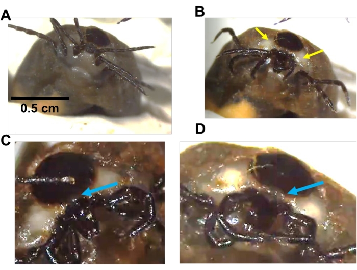
Figure 1: Manipulation of I. scapularis Gené's organ. (A) Diagram of Gene's organ. Top: Gene's organ under the scutum. Bottom: everted gland and mouthparts folded down. (B) A replete female under the microscope secured by clay. (C) A female moves her mouthparts on the ventral surface during egg-laying to extend Gene's organ. Yellow arrows show white patches and can be used as a reference for Gene's organ location. These areas are visible in females laying eggs for 2-3 weeks. (D,E) Gene's organ (a small bubble in D and extended in E) is visible between the scutum and capitulum. The horns of the Gené's organ extend as the mouthparts bend downwards (E). The blue arrow shows the location to insert a Tungsten needle to remove the gland. Please click here to view a larger version of this figure.
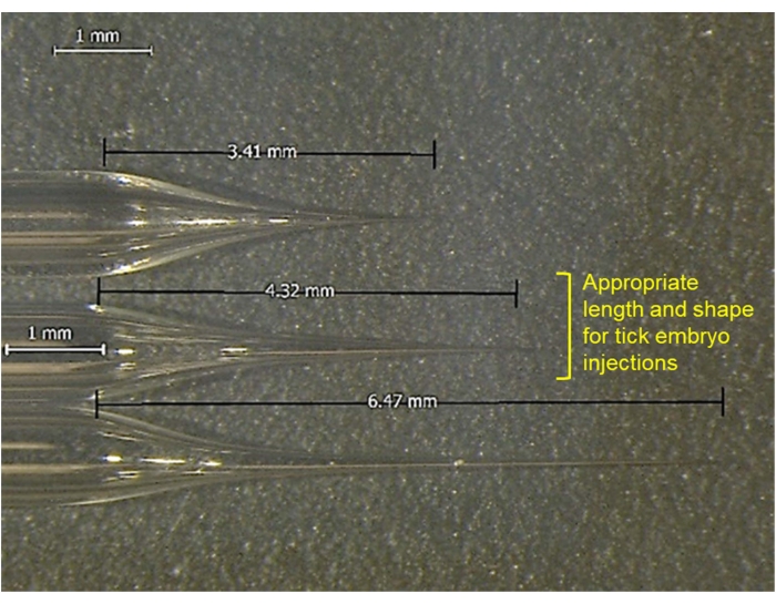
Figure 2: Comparison of the shape of glass injection needles. The middle one is used for tick embryos. Please click here to view a larger version of this figure.
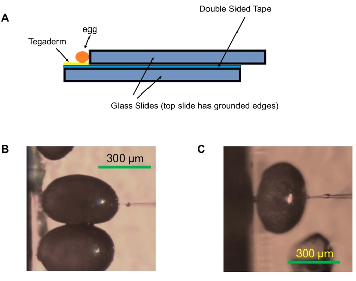
Figure 3: Egg alignment for injections. (A) Glass slide setup used for embryo injections. Microscope glass slides are attached, leaving a gap using double-sided tape, and the transparent film dressing is placed on the tape. The eggs are aligned on the edge of the slide. (B) Optimal alignment of eggs with a long axis perpendicular to the edge of the slide. (C) Less effective alignment of eggs with the long axis parallel to the edge of the slide setup. Please click here to view a larger version of this figure.
Construct injected | Time after egg-laying | Number of eggs injected | Number of larvae hatched | Survival percentage |
sgRNA + Cas9 | ≤12 h | 2,396 | 147 | 6.14 |
Gene 1 | ||||
sgRNA + Cas94 | ≤12 h | 3, 135 | 269 | 8.58 |
Gene 2 | ||||
sgRNA + Cas94 | ≤12 h | 2, 460 | 139 | 5.65 |
Gene 3 | ||||
Wolbachia | ≥ 24 h (24-36 h) | 1, 765 | 72 | 4.08 |
sgRNA + Cas9 | 48-60 h | 191 | 5 | 2.62 |
Gene 2 |
Table 1: Successful egg injection and larval hatching in Ixodes scapularis.
This is the first protocol developed to inject early tick embryos successfully. A survival rate of ~4%-8% has been achieved, which is comparable to embryo injection in other well-established insect models5.
As this is the initial protocol, it is anticipated that this protocol will be further refined and specialized to individual tick species. In particular, injection timing will vary from species to species, dependent upon embryogenesis, especially the timing of cellularization. The preliminary data suggest that I. scapularis eggs do not go through rapid nuclear division in the first 24 h after laying and cellularization occurs a few days later (unpublished data). We have already used this protocol for plasmid delivery4, CRISPR-mediated gene knockout4, and delivery of bacteria (Wolbachia) (Table 1). It is expected that this embryo injection protocol will provide opportunities to generate transgenic ticks and will make gene knockout and knock-in studies feasible in any lab. This will prove valuable for accelerating studies exploring tick biology and tick-pathogen-host interfaces.
Critical steps within the protocol
It is important to collect eggs within 24 h of laying because this protocol has been standardized for early embryos. If eggs are collected after this time, a longer treatment of benzalkonium chloride and NaCl is needed, and the survival is lower in older eggs (Table 1). The chorion hardens over time, making it difficult to do controlled desiccation for microinjections in eggs older than 24 h. It was noticed that injecting fewer eggs at a time helps with survival because the treated eggs tend to desiccate rapidly when removed from the 1% NaCl solution. If the eggs desiccate too much, they will die, but they will burst during microinjection if not desiccated appropriately. Therefore, optimizing the appropriate desiccation time is necessary for different tick species or strains. Furthermore, keeping the eggs undisturbed after microinjections in high humidity conditions is critical.
Limitations and future directions
Once the embryos are dewaxed, they tend to desiccate rapidly, so the process of aligning them on the slide and injecting them must be conducted quickly. The rapid improvement in speed and accuracy can only be achieved through constant practice and patience. Our future work will be focused on improving the survival percentage of embryos after injection and identifying the timing of germ cell formation to ensure heritable mutations.
The authors have nothing to disclose.
The authors acknowledge Channa Aluvihare and Yonus Gebermicale, ITF, UMD, for insight and support during the initial phase of protocol development. Tungsten needles were a generous gift from David O'Brochta, ITF, UMD. We are thankful to Dr. Ladislav Simo for testing this protocol in I. ricinus and for insightful discussions. This project was funded by NIH-NIAID R21AI128393 and Plymouth Hill Foundation, NY to MG-N, startup funds from the University of Nevada to AN, the National Science Foundation Grant No. 2019609 to MG-N and AN, and a Peer-to-Peer Grant from IGTRCN to AS.
Aluminum silicate capillaries, with filament | Sutter instruments | AF100-64-10 | Embryo injection |
Benzalkonium chloride 50% in water, 25 g | TCI-America | B0414 | Embryo treatment, 25 g is approximately 25 mL |
Filter paper | Whatman | 1001-090 | Post-injection care |
Forceps | Thomas Scientific | 300-101 | Gene`s organ manipulation |
Lab Wipes | Genesee Scientific | 88-115 | |
Microloader tips | Eppendorf | 930001007 | Loading the pulled needles |
Micromanipulator | Sutter instruments | ROE-200 | Embryo injection |
Microscopic slides- plain, ground edges | Genesee Scientific | 29-100 | Embryo alignment, ground edges are preferred, beveled edges could obscure the eggs from view |
NaCl | Research Products International | S23020-500.0 | Embryo treatment |
Needle Puller | Sutter Instruments | P-1000 | |
Permanent Double sided tape | Scotch | 34-8716-3417-5 | Embryo alignment |
Petri plates | Genesee Scientific | 32-107G | Post-injection care |
Tegaderm/ Transparent film dressing | 3M Healthcare | 1628 | Embryo alignment |
Tungsten needles | Fine Science Tools | 10130-10 | Gene`s organ manipulation |
Tungsten Wire | Amazon | B08DNT7ZK3 | Gene`s organ manipulation |
XenoWorks Digital Microinjector | Sutter instruments | MPC-200 | Embryo injection |