Este artigo descreve os protocolos de preparação de amostras, redução de dados e análise de dados em estudos de eco de giro de nêutrons (NSE) de membranas lipídicas. A rotulagem criteriosa de deutério de lipídios permite o acesso a diferentes dinâmicas de membrana em comprimento mesoscópico e escalas de tempo, sobre as quais ocorrem processos biológicos vitais.
É necessária uma assinatura do JoVE para visualizar este conteúdo. Faça login ou inicie seu teste gratuito.
Artigo de método
Espectroscopia de Eco de Rotação de Nêutrons como uma sonda única para dinâmicas de membrana lipídica e interações membrana-proteína
Neste artigo
Aviso de errata
Important: There has been an erratum issued for this article. View Erratum Notice
Resumo
Resumo
Bicamadas lipídicas formam a principal matriz das membranas celulares e são a principal plataforma de troca de nutrientes, interações proteína-membrana e brotamento viral, entre outros processos celulares vitais. Para uma atividade biológica eficiente, as membranas celulares devem ser rígidas o suficiente para manter a integridade da célula e seus compartimentos, mas fluidas o suficiente para permitir que componentes de membrana, como proteínas e domínios funcionais, difundam e interajam. Este delicado equilíbrio de propriedades de membrana elástica e fluida, e seu impacto na função biológica, necessitam de uma melhor compreensão da dinâmica da membrana coletiva sobre o comprimento mesoscópico e escalas de tempo dos principais processos biológicos, por exemplo, deformações de membrana e eventos de ligação de proteínas. Entre as técnicas que podem efetivamente sondar esse alcance dinâmico está a espectroscopia de nêutrons spin echo (NSE). Combinado com a rotulagem de deutério, o NSE pode ser usado para acessar diretamente flutuações de dobra e espessura, bem como dinâmica mesoscópica de características de membrana selecionadas. Este artigo fornece uma breve descrição da técnica NSE e descreve os procedimentos para a realização de experimentos NSE em membranas liposômicas, incluindo detalhes de esquemas de preparação e deuteração da amostra, juntamente com instruções para coleta e redução de dados. O artigo também introduz métodos de análise de dados usados para extrair parâmetros de membrana chave, como o módulo de rigidez de dobra, o módulo de compressão da área e a viscosidade no plano. Para ilustrar a importância biológica dos estudos da NSE, são discutidos exemplos selecionados de fenômenos de membrana sondados pela NSE, ou seja, o efeito dos aditivos sobre a rigidez de dobra de membrana, o impacto da formação de domínios sobre as flutuações da membrana e a assinatura dinâmica das interações membrana-proteína.
Introdução
A compreensão das membranas celulares e sua função evoluiu notavelmente nas últimas décadas. A visão anterior das membranas celulares como bicamadas lipídicas passivas que definem limites celulares e proteínas de membranadoméstica 1 gradualmente se transformou em um modelo dinâmico no qual bicamadas lipídicas desempenham um papel importante na regulação de processos biológicos vitais, incluindo sinalização celular, troca molecular e função proteica — para citar alguns2,3,4,5,6
Acesso restrito. Inicie sessão ou comece um teste para visualizar este conteúdo.
Protocolo
1. Esquema de desauteização necessário para o experimento
- Para medir a flutuação de dobra, faça lipossomos totalmente protiados em D2O (D 99,9%) ou D2O-buffer (por exemplo, tampão fosfato preparado com D2O em vez de H2O). Use DMPC totalmente protiado (C36H72NO8P) e DSPC (C44H88NO8P) com
 133,4 mgs, onde XDMPC e XDSPC são as frações moles de DMPC e DSPC, aqui definidas para 0,7 e 0,3, respectivamente, e MwDM....
Acesso restrito. Inicie sessão ou comece um teste para visualizar este conteúdo.
Resultados
Estudos NSE acessando flutuações de dobra são normalmente realizados ao longo de uma faixa Q de ~ (0,04 - 0,2) Å-1. Esta faixa Q corresponde a escalas de comprimento intermediário entre a espessura da membrana e o raio lipossômico, onde a dinâmica de dobra domina. A medição sobre um intervalo Q estendido pode dar acesso a modos dinâmicos adicionais, incluindo difusão liposômica e dinâmica intramembrana. Para obter mais detalhes sobre o cruzamento na dinâmica da membrana acessada pela NSE, consulte estas public.......
Acesso restrito. Inicie sessão ou comece um teste para visualizar este conteúdo.
Discussão
NSE é uma técnica poderosa e única na medição da dinâmica mesoscópica das membranas lipídicas sob várias condições. A utilização efetiva da NSE depende da qualidade da amostra, do contraste de nêutrons e da gama de dinâmicas acessíveis que podem ser sondadas para uma determinada amostra. Assim, várias etapas críticas são necessárias para a realização de experimentos NSE bem-sucedidos e a coleta de dados de alta qualidade. Um passo fundamental para garantir o uso efetivo do tempo de feixe de nêutrons durante um experiment.......
Acesso restrito. Inicie sessão ou comece um teste para visualizar este conteúdo.
Divulgações
Os autores não declaram conflitos de interesse e não têm nada a revelar.
Agradecimentos
R. Ashkar agradece M. Nagao, L.-R. Stingaciu e P. Zolnierczuk para muitas discussões úteis e por sua assistência frequente com experimentos NSE em suas respectivas linhas de trave. Os autores reconhecem o uso de espectrômetros de eco de spin de nêutrons no NIST e ORNL. O espectrômetro NSE no NIST é apoiado pelo Centro de Dispersão de Nêutrons de Alta Resolução, uma parceria entre o Instituto Nacional de Padrões e Tecnologia e a Fundação Nacional de Ciência sob o acordo nº. DMR-1508249. O espectrômetro NSE na Fonte de Nêutrons spallation da ORNL é apoiado pela Divisão de Instalações de Usuários Científicos, Escritório de Ciências Básicas de Energia, Departamento de Ene....
Acesso restrito. Inicie sessão ou comece um teste para visualizar este conteúdo.
Materiais
| Nome | Empresa | Número de catálogo | Comentários |
|---|---|---|---|
| Clorofórmio (grau biotecnológico) | Sigma Aldrich | 496189 | Biotech. grau, ≥ 99,8%, contém 0,5-1,0% de etanol como estabilizador |
| Banho-maria circulante | Julabo | SE-12 | Circulador de aquecimento com bomba inteligente, configurações de temperatura programáveis e conexão de sensor externo para medição e controle |
| Óxido de deutério | Cambridge Isotopes Laboratories | DLM-4 | Água deuterada; Água pesada (D2O) (D, 99,9%) |
| Semimicrobalança Digital | Mettler Toledo | MS105 | Balança semimicro com capacidade de 120 g, resolução de 0,01 mg, célula de pesagem de alta resolução, portas ergonômicas e aplicação de verificação de pipetas |
| Etanol (grau de biologia molecular) | Sigma Aldrich | E7023 | Etanol à prova de 200 para aplicações de biologia molecular |
| Pipetas de vidro | VWR | 36360-536 | Pipetas de Pasteur de vidro de cal sodada |
| frascos de vidro | Thermo Scientific | B7990-1 | Frascos de vidro de borosilicato com tampas de septo de PTFE/Silione |
| Freezer de grau de laboratório | Fisher Scientific | IU2886D | Freezer de temperatura ultrabaixa (-86 a -50 C) para armazenamento a longo prazo de lipídios e proteínas |
| Lipídios (produzidos ou perdeuterados) | Os lipídios polares | Avanti variam de acordo com o lipídio | Os lipídios podem ser adquiridos da Avanti em pó ou em uma solução de clorofórmio com as quantidades e esquemas de deuteração necessários. |
| Purificador de água | Millipore Sigma | ZRQSVP3US | Direct-Q® 3 Sistemas de Purificação de Água UV que fornecem água pura e ultrapura com uma lâmpada UV embutida para reduzir os níveis de orgânicos para produtos biológicos aplicações |
| Mini Conjunto de Extrusora | Avanti Polar Lipids | 610020 | O conjunto de mini-extrusora inclui mini-extrusora, bloco de aquecimento, 2 seringas GasTight, e 2 O-rings, membranas de policarbonato e suportes de filtro |
| Conexões de conexão rápida | Grainger | 2YDA1 e 2YDA7 | Conexões de tubo de botão de pressão para aplicações de circulação de água QuickConnect, por exemplo, extrusão de vesículas de alta temperatura |
| Bomba | de seringaSyringePump.com | New Era-1000 Bomba de | seringa totalmente programável para infusão e retirada; programa até 41 fases de bombeamento com taxas de bombeamento ajustáveis, volumes dispensados e ciclos de extrusão |
| Banho ultra-sónico | Fisher Scientific | CPX2800 | Banho ultra-sónico com controlo de temperatura com funcionalidade programável para aplicações de desgaseificação e ultra-sons |
| Forno a vácuo | Thermo Scientific | 3608 | Forno a vácuo de 0,7 pés cúbicos com termostato de limite alto embutido protege contra superaquecimento |
| Misturador de Vórtice | Fisher Scientific | 02-215-414 | Controle analógico de velocidade variável que permite partida em baixa rotação para agitação suave ou mistura de alta velocidade para vórtice vigoroso de amostras |
Referências
- Singer, S. J., Nicolson, G. L. The fluid mosaic model of the structure of cell membranes. Science. 175 (4023), 720-731 (1972).
- Andersen, O. S., Koeppe, R. E. Bilayer thickness and membrane protein function: an energetic perspective. Annual ....
Acesso restrito. Inicie sessão ou comece um teste para visualizar este conteúdo.
Reimpressões e permissões
Errata
Formal Correction: Erratum: Neutron Spin Echo Spectroscopy as a Unique Probe for Lipid Membrane Dynamics and Membrane-Protein Interactions
Posted by JoVE Editors on 8/06/2021. Citeable Link.
An erratum was issued for: Neutron Spin Echo Spectroscopy as a Unique Probe for Lipid Membrane Dynamics and Membrane-Protein Interactions. The Introduction, Protocol, and Representative Results sections have been updated.
In the Introduction, the fith pargraph was updated from:
Besides direct access to the length and time scale of membrane dynamics, NSE has the inherent capabilities of neutron isotope sensitivity52. Specifically, the ability of neutrons to interact differently with the isotopes of hydrogen, the most abundant element in biological systems, results in a different neutron scattering length density,34 or NSLD (the equivalent of the optical index of refraction50), when protium is substituted by deuterium. This enables an approach known as contrast variation, which is commonly used to highlight specific membrane features or conceal others — the latter scenario is referred to as contrast matching. A frequent application of contrast variation/matching is the substitution of water (NSLD = -0.56 × 10⁻⁶ Å-2) by heavy water or D2O (NSLD = 6.4 × 10⁻⁶ Å-2) to amplify the neutron signal from protiated lipid membranes (NSLD ~ 2 × 10⁻⁶ Å-2). This approach is highly effective in studies of membrane structure because the penetration of D2O into the headgroup region of the membrane allows accurate determination of the membrane thicknesses (see Figure 2A, left panel) and of the location of different lipid subgroups when more sophisticated models are applied53,54. This paper highlights some examples on the use of contrast variation for studies of collective dynamics in biomimetic membranes and select membrane features.
to:
Besides direct access to the length and time scale of membrane dynamics, NSE has the inherent capabilities of neutron isotope sensitivity52. Specifically, the ability of neutrons to interact differently with the isotopes of hydrogen, the most abundant element in biological systems, results in a different neutron scattering length density,34 or NSLD (the equivalent of the optical index of refraction50), when protium is substituted by deuterium. This enables an approach known as contrast variation, which is commonly used to highlight specific membrane features or conceal others — the latter scenario is referred to as contrast matching. A frequent application of contrast variation/matching is the substitution of water (NSLD = -0.56 × 10⁻⁶ Å-2) by heavy water or D2O (NSLD = 6.4 × 10⁻⁶ Å-2) to amplify the neutron signal from protiated lipid membranes (NSLD ~ 0 × 10-6 Å-2). This approach is highly effective in studies of membrane structure because the penetration of D2O into the headgroup region of the membrane allows accurate determination of the membrane thicknesses (see Figure 2A, left panel) and of the location of different lipid subgroups when more sophisticated models are applied53,54. This paper highlights some examples on the use of contrast variation for studies of collective dynamics in biomimetic membranes and select membrane features.
In the Protocol, step 1.1 was updated from:
For bending fluctuation measurements, make fully protiated liposomes in D2O (D 99.9%) or D2O-buffer (e.g., phosphate buffer prepared with D2O instead of H2O). Use fully protiated DMPC (C36H72NO8P) and DSPC (C44H88NO8P) with 133.4 mg, where XDMPC and XDSPC are the mole fractions of DMPC and DSPC, here set to 0.7 and 0.3, respectively, and MwDMPC and MwDSPC are the molar weights given by 677.9 g/mol and 790.1 g/mol, respectively. Similarly, mDSPC = 66.6 mg. This deuteration scheme increases the scattering contrast between the membrane (NSLD ~ 2 × 10⁻⁶ Å-2) and the deuterated buffer (NSLD ~ 6.4 × 10⁻⁶ Å-2) and amplifies the signal from membrane undulations (see Figure 2A left panel).
to:
For bending fluctuation measurements, make fully protiated liposomes in D2O (D 99.9%) or D2O-buffer (e.g., phosphate buffer prepared with D2O instead of H2O). Use fully protiated DMPC (C36H72NO8P) and DSPC (C44H88NO8P) with 133.4 mg, where XDMPC and XDSPC are the mole fractions of DMPC and DSPC, here set to 0.7 and 0.3, respectively, and MwDMPC and MwDSPC are the molar weights given by 677.9 g/mol and 790.1 g/mol, respectively. Similarly, mDSPC = 66.6 mg. This deuteration scheme increases the scattering contrast between the membrane (NSLD ~ 0 × 10-6 Å-2) and the deuterated buffer (NSLD ~ 6.4 × 10⁻⁶ Å-2) and amplifies the signal from membrane undulations (see Figure 2A left panel).
In the Representative Results, the fist pagargaph was updted from:
NSE studies accessing bending fluctuations are typically performed over a Q-range of ~ (0.04 - 0.2) Å-1. This Q-range corresponds to intermediate length scales between the membrane thickness and the liposomal radius, where bending dynamics dominate. Measurement over an extended Q-range can give access to additional dynamic modes, including liposomal diffusion and intramembrane dynamics. For more details on the cross-over in membrane dynamics accessed by NSE, check these relevant publications25,71. It is important to emphasize that NSE signals are proportional to:  , where Icoh and Iinc are, respectively, the coherent and incoherent scattering intensity from the sample. Therefore, it is advisable to prepare NSE liposomal samples in deuterated buffers (i.e., buffers prepared with D2O instead of H2O) to minimize the incoherent scattering signal, mainly contributed by the hydrogen content of the sample. However, in some cases intermediate deuteration schemes (i.e., using mixtures of D2O and H2O) might be necessary to obtain optimal contrast conditions. Typically, NSE measurements of membrane bending fluctuations are performed on fully protiated liposomes in deuterated buffer, referred to as fully contrasted liposomes in Figure 5. This deuteration scheme results in a large NSLD difference between the membrane core (~2 × 10⁻⁶ Å-2) and its deuterated fluid environment (~6.4 × 10⁻⁶ Å-2), which significantly enhances the scattering signal from the liposomal membranes and improves the measurement statistics of bending dynamics. This contrast scheme (Figure 2A left panel) is frequently utilized in studies of bending rigidity of lipid membranes with single38,72 and multiple39,66 lipid components and in studies of membrane softening/stiffening by biological inclusions (e.g., cholesterol, drug molecules, peptides/proteins)36,37,73,74,75, and synthetic additives (e.g., nanoparticles)76,77.
to:
NSE studies accessing bending fluctuations are typically performed over a Q-range of ~ (0.04 - 0.2) Å-1. This Q-range corresponds to intermediate length scales between the membrane thickness and the liposomal radius, where bending dynamics dominate. Measurement over an extended Q-range can give access to additional dynamic modes, including liposomal diffusion and intramembrane dynamics. For more details on the cross-over in membrane dynamics accessed by NSE, check these relevant publications25,71. It is important to emphasize that NSE signals are proportional to: , where Icoh and Iinc are, respectively, the coherent and incoherent scattering intensity from the sample. Therefore, it is advisable to prepare NSE liposomal samples in deuterated buffers (i.e., buffers prepared with D2O instead of H2O) to minimize the incoherent scattering signal, mainly contributed by the hydrogen content of the sample. However, in some cases intermediate deuteration schemes (i.e., using mixtures of D2O and H2O) might be necessary to obtain optimal contrast conditions. Typically, NSE measurements of membrane bending fluctuations are performed on fully protiated liposomes in deuterated buffer, referred to as fully contrasted liposomes in Figure 5. This deuteration scheme results in a large NSLD difference between the membrane core (~0 × 10-6 Å-2) and its deuterated fluid environment (~6.4 × 10⁻⁶ Å-2), which significantly enhances the scattering signal from the liposomal membranes and improves the measurement statistics of bending dynamics. This contrast scheme (Figure 2A left panel) is frequently utilized in studies of bending rigidity of lipid membranes with single38,72 and multiple39,66 lipid components and in studies of membrane softening/stiffening by biological inclusions (e.g., cholesterol, drug molecules, peptides/proteins)36,37,73,74,75, and synthetic additives (e.g., nanoparticles)76,77.
In the Representative Reults, Figure 2 was updated from:

Figure 2: Examples of possible deuteration schemes in NSE experiments on lipid membranes. (A) Left: Fully contrasted membranes, e.g., protiated membranes in deuterated buffer, showing the NSLD profile along the normal to the membrane surface. The difference in the NSLD between the headgroup (~2 × 10⁻² Å-2) and tail region (~4.5 × 10⁻⁶ Å-2) of the membrane is due to the headgroup hydration with deuterated buffer. Right: Tail-contrast matched membranes such that the hydrocarbon tail region of the membrane has the same NSLD as the buffer, as shown in the corresponding NSLD profile along the membrane normal. (B) Domain-forming membranes with two neutron contrast schemes where the domains (center) or the matrix (left) are contrast-matched to the buffer, enabling selective studies of matrix or domain dynamics, respectively. This figure has been modified from Nickels et al., JACS 201541. (C) Asymmetric membranes prepared by cyclodextrin exchange between protiated and deuterated lipid vesicles, resulting in the deuteration of one membrane leaflet while keeping the other leaflet protiated. This allows studies of the bending dynamics of the protiated leaflet and provides insights into the mechanical coupling between opposing leaflets in asymmetric membranes. This figure has been modified from Rickeard et al., Nanoscale 202040. Please click here to view a larger version of this figure.
to:
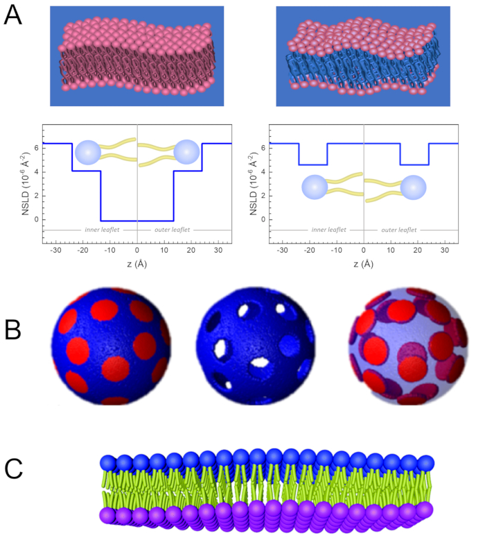
Figure 2: Examples of possible deuteration schemes in NSE experiments on lipid membranes. (A) Left: Fully contrasted membranes, e.g., protiated membranes in deuterated buffer, showing the NSLD profile along the normal to the membrane surface. The difference in the NSLD between the tail region (~0 × 10-2 Å-2) and headgroup region (~4.5 × 10⁻⁶ Å-2) of the membrane is due to the headgroup hydration with deuterated buffer. Right: Tail-contrast matched membranes such that the hydrocarbon tail region of the membrane has the same NSLD as the buffer, as shown in the corresponding NSLD profile along the membrane normal. (B) Domain-forming membranes with two neutron contrast schemes where the domains (center) or the matrix (left) are contrast-matched to the buffer, enabling selective studies of matrix or domain dynamics, respectively. This figure has been modified from Nickels et al., JACS 201541. (C) Asymmetric membranes prepared by cyclodextrin exchange between protiated and deuterated lipid vesicles, resulting in the deuteration of one membrane leaflet while keeping the other leaflet protiated. This allows studies of the bending dynamics of the protiated leaflet and provides insights into the mechanical coupling between opposing leaflets in asymmetric membranes. This figure has been modified from Rickeard et al., Nanoscale 202040. Please click here to view a larger version of this figure.
