Summary

タンパク質と水和水のピコ秒ナノ秒ダイナミクスを研究するための高分解能中性子分光法

Published: April 28, 2022
doi:

Summary

中性子後方散乱分光法は、タンパク質とその水和水のps-nsダイナミクスへの非破壊でラベルフリーのアクセスを提供します。このワークフローには、アミロイドタンパク質に関する2つの研究、すなわち凝集中のリゾチームの時間分解ダイナミクスと、繊維形成時のタウの水和水ダイナミクスに関する研究が提示されています。

Abstract

中性子散乱は、重水素以外の標識なしに、非破壊的な方法で広範囲のエネルギーのサンプル内のダイナミクスをプローブする可能性を提供します。特に、中性子後方散乱分光法は、複数の散乱角度で同時に散乱信号を記録し、ps-nsタイムスケールでの生物系のダイナミクスの研究に適しています。D2Oおよび場合によっては重水素化緩衝液成分を使用することにより、この方法は、液体状態のタンパク質の重心拡散と骨格および側鎖運動(内部ダイナミクス)の両方をモニタリングすることを可能にする。

さらに、水和水の動態は、H2Oで水和された過重水素化タンパク質の粉末を使用することによって研究することができる。この論文では、タンパク質と水分補給の水動態を調査するために、ラウエランジュバン研究所(ILL)の装置IN16Bで採用されたワークフローを紹介します。蒸気交換を用いた溶液サンプルおよび水和タンパク質粉末サンプルの調製について説明する。タンパク質と水和水ダイナミクスの両方のデータ解析手順は、中性子後方散乱分光計で取得できるさまざまなタイプのデータセット(準弾性スペクトルまたは固定ウィンドウスキャン)について説明されています。

この方法は、アミロイドタンパク質を含む2つの研究で示されています。リゾチームのμmサイズの球状凝集体(微粒子と表記)への凝集は、IN16Bで調査された空間および時間範囲でのワンステッププロセスで発生することが示されていますが、内部ダイナミクスは変化しません。さらに、タウの水和水の動態を過重水素化タンパク質の水和粉末で研究した。水の並進運動はアミロイド線維の形成時に活性化されることが示されている。最後に、他の実験的生物物理学的方法に関するダイナミクスの研究に関して中性子散乱がどのように位置付けられているかに関して、プロトコルの重要なステップが議論されます。

Introduction

中性子は電荷のない巨大な粒子であり、基礎物理学から生物学まで、さまざまな分野のサンプルを調べるために長年にわたって成功裏に使用されてきました1。生物学的用途では、小角中性子散乱、非弾性中性子散乱、中性子結晶構造解析および反射率測定が広く使用されています2,3,4非弾性中性子散乱は、特定の標識自体を必要とせずにダイナミクスのアンサンブル平均測定を提供しサイズやタンパク質に依存しない信号品質を提供します5。測定は、重水素化細菌ライセートやin vivoなどの細胞内培地を模倣する研究中のタンパク質の非常に複雑な環境を使用して行うことができます3,6,7ダイナミクス、すなわち、i)飛行時間によるサブps-psダイナミクスへのアクセス、ii)後方散乱によるps-nsダイナミクスへのアクセス、iii)nsから数百nsまでのダイナミクスへのアクセスなど、さまざまな実験セットアップを使用してダイナミクスを研究することができます。中性子後方散乱は、ブラッグの法則2d sinθ = nλを利用しており、dは結晶内の平面間の距離、θは散乱角、nは散乱次数、λは波長です。検出器への後方散乱に結晶を使用することで、エネルギーの高分解能(通常~0.8μeV)を達成することができます。エネルギー交換を測定するには、後方散乱で結晶を運ぶドップラードライブを使用して入射中性子波長8910を定義および調整するか(図1)、エネルギー分解能の低下を犠牲にして飛行時間型セットアップを使用できます11

Figure 1
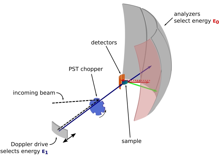
図1:ドップラー駆動の中性子後方散乱分光計のスケッチ。入射ビームは位相空間変換(PST)チョッパ42に当たり、試料位置における磁束を増加させる。次に、ドップラードライブによってサンプルに向かって後方散乱され、ドップラードライブはエネルギーE1(シアンの矢印)を選択します。次に、中性子はサンプルによって散乱され(矢印の色で表される異なるエネルギーで)、Si 111結晶で作られた分析装置は、特定のエネルギーE0(ここでは赤い色の矢印)の中性子のみを後方散乱します。したがって、運動量移動qは検出器アレイ上の中性子の検出位置から得られ、エネルギー移動は差E1-E0から得られます。PSTによって生成された中性子パルスに予想される飛行時間は、検出器管に向かって直接散乱された中性子からの信号を破棄するために使用されます。略語:PST =位相空間変換。この図の拡大版を表示するには、ここをクリックしてください。

後方散乱分光法では、タンパク質などの水素プロトンに富むサンプルからのシグナルへの主な寄与は、散乱強度Sinc(q、ω)が式(1)12で示されるインコヒーレント散乱から来ています
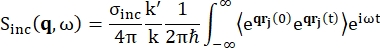
Equation 1 (1)

ここでσincは考慮される元素のインコヒーレント断面であり、k’は散乱波ベクトルのノルム、kは入射波ベクトルのノルム、q(= k – k’)運動量移動、r j(t)時間tにおける原子jの位置ベクトル、ωは入射中性子と系との間のエネルギー移動に対応する周波数である。角括弧はアンサンブル平均を示します。したがって、インコヒーレント散乱は、原子位置のアンサンブル平均単粒子自己相関を時間とプローブし、システム内のすべての原子と異なる時間起源(アンサンブル平均)で平均化された自己ダイナミクスを提供します。散乱関数は、中間散乱関数 I(q, t) のときのフーリエ変換であり、式 (2) で示されるファンホーブ相関関数の空間でのフーリエ変換と見なすことができます。
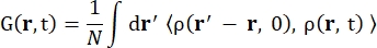
Equation 2 (2)

ここで、ρ(r,t)は、位置rと時間t13で原子を見つける確率密度です。

フィッキアン拡散過程の場合、自己拡散関数は、γ = Dq2で与えられる線幅のローレンツ分布からなる散乱関数の二重フーリエ変換の後に生じます(式(3)を参照)。
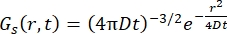
Equation 10 (3)

より洗練されたモデルが開発され、ps-ns内部タンパク質ダイナミクス14のSingwiとSjölanderによるジャンプ拡散モデルや、水和水15,16,17のSearsによる回転モデルなど、有用であることがわかりました。

フランスのグルノーブルにあるILLの中性子後方散乱(NBS)装置IN16B 8,9(補足図S1)では、タンパク質で一般的に使用されるセットアップは、入射波長を調整するためのドップラードライブを備えたアナライザー用のSi 111結晶で構成され(補足図S2A)、それによって運動量伝達範囲~0.2 Å-1 < q < ~2 Å-1および30 μeVのエネルギー伝達範囲にアクセスできます< Equation 3< 30μeV-数psから数nsの範囲のタイムスケールと数Åの距離に対応します。さらに、IN16Bは、固定エネルギー伝達でのデータ収集を含む弾性および非弾性固定ウィンドウスキャン(E/IFWS)10を実行する可能性を提供します。中性子を扱う場合、磁束が制限されるため、E/IFWSは1回のエネルギー伝達で磁束を最大化できるため、満足のいく信号対雑音比を得るために必要な取得時間を短縮できます。より最近のオプションは、後方散乱および飛行時間型分光計(BATS)モード11であり、ドップラー駆動よりも高い磁束で、広範囲のエネルギー移動(例えば、-150μeV<< Equation 3 150μeV)の測定を可能にしますが、エネルギー分解能は低くなります(補足図S2B)。

中性子散乱の重要な特性は、インコヒーレント断面積σincが水素に対して重水素よりも40倍高い値を持ち、生物学的サンプルに一般的に見られる他の元素に対しては無視できることです。したがって、液体環境におけるタンパク質の動態は、重水素化緩衝液を用いて研究することができ、粉末状態は、D2Oで水和した水素化タンパク質粉末によるタンパク質内部動態の研究、またはH2Oで水和した過重化タンパク質粉末の水和水の研究を可能にする。液体状態では、中性子後方散乱は通常、タンパク質の重心自己拡散(フィッキアン型拡散)とその内部ダイナミクスに同時にアクセスすることができます。後者は、通常、いわゆるジャンプ拡散モデルなどによって記述されるバックボーンおよびサイドチェーン運動です3,18。水素化タンパク質粉末では、タンパク質の拡散は存在せず、内部ダイナミクスのみをモデル化する必要があります。水和水の場合、水分子の並進運動および回転運動の寄与は、運動量移動qに対して異なる依存性を示し、これは、データ分析プロセスにおけるそれらの区別を可能にする17

この論文は、β鎖のスタックからなる標準的な形に展開し、凝集し、いわゆるクロスβパターン19,20-そして細長い繊維を形成することができることが判明したタンパク質の研究による中性子後方散乱法を示しています。これはいわゆるアミロイド凝集であり、アルツハイマー病やパーキンソン病などの神経変性疾患における中心的な役割のために広く研究されています21,22。アミロイドタンパク質の研究は、それらが果たすことができる機能的役割23,24または新規生体材料の開発のためのそれらの高い可能性によっても動機付けられています25アミロイド凝集の物理化学的決定要因は依然として不明であり、過去数年間の驚異的な進歩にもかかわらず、アミロイド凝集の一般理論は利用できません21,26

アミロイド凝集は、タンパク質の構造と経時的な安定性の変化を意味し、その研究は当然、タンパク質の立体構造安定性、タンパク質機能、およびタンパク質エネルギーランドスケープに関連するダイナミクスを意味します27。ダイナミクスは、最速の運動28のエントロピー寄与を通じて特定の状態の安定性に直接関連しており、タンパク質の機能は、光感受性タンパク質29 のサブpsからドメイン運動のmsまで、ピコ秒-ナノ秒のダイナミクス30によって促進されるさまざまなタイムスケールの運動によって維持できます。

中性子後方散乱分光法を使用してアミロイドタンパク質を研究する2つの例、1つはタンパク質のダイナミクスを研究するための液体状態、もう1つは水和水のダイナミクスを研究するための水和粉末状態の2つの例を紹介します。第1の例は、リゾチームのμmサイズの球体(微粒子と呼ばれる)への凝集に関するものであり、続いてリアルタイムで5、第2の例は、ヒトタンパク質タウ31の天然状態および凝集状態における水動態の比較に関するものである。

リゾチームは免疫防御に関与する酵素で、129個のアミノ酸残基で構成されています。リゾチームは、pD10.5および90°Cの温度で重水素化緩衝液中で微粒子を形成することができます。 中性子散乱により、リゾチームの重心拡散係数の時間発展がチオフラビンT蛍光(アミロイド交差βパターンの形成をモニターするために使用される蛍光プローブ32)の単一の指数関数的速度論に従うことを示し、粒子形成上部構造と交差βパターンが同じ速度で1つのステップで発生することを示しています。さらに、内部ダイナミクスは凝集プロセス全体を通して一定であり、これはNBS装置では観察できない速い立体構造変化、または凝集時にタンパク質の内部エネルギーに大きな変化がないことによって説明できます。

ヒトタンパク質タウは、いわゆる2N4Rアイソフォームの441アミノ酸からなる天然変性タンパク質(IDP)であり、アルツハイマー病に特に関与しています33。過重水素化タンパク質タウの粉末に対する中性子後方散乱を用いて、繊維状態で水和水ダイナミクスが増加し、並進運動を受ける水分子の集団が多いことを示しました。この結果は、水和水エントロピーの増加がタウのアミロイド細動を引き起こす可能性があることを示唆している。

Protocol

1. 液体状態のタンパク質用の重水素化バッファーを調製します 緩衝液のすべての成分を純粋なD2Oに溶解する。 pH電極をH2Oで較正した場合、NaODまたはDCl34を用いて式pD = pH + 0.4に従ってpDを調整する。注:H2Oの代わりにD2Oを使用すると、タンパク質の溶解度に影響を与える可能性があり、緩衝条件を適応させる必要?…

Representative Results

リゾチームの微粒子への凝集は、重水素化緩衝液(pD 10.5で0.1 M NaCl)中で50 mg/mLのタンパク質濃度で90°Cで実施しました。微粒子の形成は、90°Cへの温度上昇によって引き起こされ、6時間以内に発生します(補足図S8)。データ取得は、上記のプロトコルで説明されているように、IN16Bで実行されました(データはILLによって永続的にキュレーションされ、http://dx.doi.org/10.5291/ILL-DATA.8-04-811 …

Discussion

中性子分光法は、重水素化を使用した場合のタンパク質のサイズや溶液の複雑さに関係なく、タンパク質サンプルのアンサンブル平均ps-nsダイナミクスをプローブできる唯一の方法です6。具体的には、溶液中でのタンパク質集合体の自己拡散を調べることにより、そのような集合体の流体力学的サイズを明確に決定することができる。それにもかかわらず、この方法は一般?…

Disclosures

The authors have nothing to disclose.

Acknowledgements

著者らは、ドイツのガルヒングにあるハインツ・マイヤー・ライプニッツ・ツェントルムにあるユーリッヒ中性子科学センターのミカエラ・ザンポーニ氏に、SPHERES装置で行われた中性子散乱実験の一部に感謝している。この研究は、欧州連合がHPRI-2001-50065およびRII3-CT-2003-505925の契約に基づいて資金提供する重水素化研究所(DLAB)コンソーシアムの活動、および英国工学物理科学研究評議会(EPSRC)が資金提供する助成金GR/R99393/01およびEP/C015452/1に基づくラウエランジュバンEMBL研究所内の活動から恩恵を受けています。主要行動を通じた第7次枠組みプログラムに基づく欧州委員会による支援:欧州研究領域、研究インフラストラクチャの強化が認められています[契約226507(NMI3)]。ケビン・ポウノットとクリスチャン・ベックは、ポスドクフェローシップの資金提供について、連邦教育研究省(BMBF、助成金番号05K19VTB)に感謝します。

Materials

Aluminum sample holder Not commercially available. Either the local contact on the instrument can provide them or they can be manufactured based on a technical drawing that can be provided by the local contact.
Deuterium chloride, 35 wt. % in D2O, ≥99 atom % D Sigma-Aldrich
543047
Deuterium oxide (D, 99.9%) Eurisotop DLM-4DR-PK
Dow Corning high-vacuum silicone grease Sigma-Aldrich Z273554-1EA
Ethanol 96%, EMSURE Reag. Ph Eur Sigma-Aldrich 1.5901
Glass dessicator VWR   75871-660
Glass dessicator plate, 140 mm VWR 89038-068
Indium wire, 1.0 mm (0.04 in) dia, Puratronic, 99.999% Alfa Aesar 00470.G1
Lysozyme from chicken egg white dialyzed, lyophilized, powder, ~100,000 U/mg Sigma-Aldrich 62970
nPDyn v3.x see github.com/kpounot/nPDyn, model functions fot fitting also included in the software
OHAUS AX324 Adventurer balance, internal calibration Dutscher 92641
Phosphorus pentoxide, ReagentPlus, 99% Sigma-Aldrich 214701
Pipette ErgoOne 0.5-10 μL Starlab S7100-0510
Pipette ErgoOne 100-1,000 μL Starlab S7100-1000
Pipette ErgoOne 20-200 μL Starlab S7100-2200
Pipette tip TipOne 1,000 μL Starlab S1111-6001
Pipette tip TipOne 10 μL Starlab S1111-3200
Pipette tip TipOne 200 μL Starlab S1111-0206
Sodium deuteroxide solution, 40 wt. % in D2O, 99.5 atom % D Sigma-Aldrich 372072

References

  1. Jacrot, B. Des neutrons pour la science: Histoire de l’Institut Laue-Langevin. Des neutrons pour la science. EDP Sciences. , (2021).
  2. Mahieu, E., Gabel, F. Biological small-angle neutron scattering: recent results and development. Acta Crystallographica Section D. 74 (8), 715-726 (2018).
  3. Grimaldo, M., Roosen-Runge, F., Zhang, F., Schreiber, F., Seydel, T. Dynamics of proteins in solution. Quarterly Reviews of Biophysics. 52, 7 (2019).
  4. Martel, A., et al. Membrane permeation versus amyloidogenicity: A multitechnique study of islet amyloid polypeptide interaction with model membranes. Journal of the American Chemical Society. 139 (1), 137-148 (2017).
  5. Pounot, K., et al. Tracking internal and global diffusive dynamics during protein aggregation by high-resolution neutron spectroscopy. The Journal of Physical Chemistry Letters. 11 (15), 6299-6304 (2020).
  6. Grimaldo, M., et al. Protein short-time diffusion in a naturally crowded environment. The Journal of Physical Chemistry Letters. 10 (8), 1709-1715 (2019).
  7. Jasnin, M., Stadler, A., Tehei, M., Zaccai, G. Specific cellular water dynamics observed in vivo by neutron scattering and NMR. Physical Chemistry Chemical Physics. 12 (35), 10154-10160 (2010).
  8. Frick, B. The neutron backscattering spectrometer IN16 at ILL-high energy resolution with high intensity and excellent signal-to-noise ratio. Neutron News. 13 (2), 15-22 (2002).
  9. Frick, B., Mamontov, E., van Eijck, L., Seydel, T. Recent backscattering instrument developments at the ILL and SNS. Zeitschrift für Physikalische Chemie. 224 (1-2), 33-60 (2010).
  10. Frick, B., Combet, J., van Eijck, L. New possibilities with inelastic fixed window scans and linear motor Doppler drives on high resolution neutron backscattering spectrometers. Nuclear Instruments and Methods in Physics Research Section A: Accelerators, Spectrometers, Detectors and Associated Equipment. 669, 7-13 (2012).
  11. Appel, M., Frick, B., Magerl, A. A flexible high speed pulse chopper system for an inverted neutron time-of-flight option on backscattering spectrometers. Scientific Reports. 8 (1), 13580 (2018).
  12. Squires, G. L. . Introduction to the theory of thermal neutron scattering. , (1996).
  13. Singwi, K. S., Sjölander, A. Diffusive motions in water and cold neutron scattering. Physical Review. 119 (3), 863-871 (1960).
  14. Sears, V. F. Theory of cold neutron scattering by homonuclear diatomic liquids: i. free rotation. Canadian Journal of Physics. 44 (6), 1279-1297 (1966).
  15. Sears, V. F. Theory of cold neutron scattering by homonuclear liquid: ii. hindered rotation. Canadian Journal of Physics. 44 (6), 1299-1311 (1966).
  16. Schirò, G., et al. Translational diffusion of hydration water correlates with functional motions in folded and intrinsically disordered proteins. Nature Communications. 6, 6490 (2015).
  17. Grimaldo, M., et al. Hierarchical molecular dynamics of bovine serum albumin in concentrated aqueous solution below and above thermal denaturation. Physical Chemistry Chemical Physics. 17 (6), 4645-4655 (2015).
  18. Eanes, E. D., Glenner, G. G. X-ray diffraction studies on amyloid filaments. Journal of Histochemistry & Cytochemistry. 16 (11), 673-677 (1968).
  19. Bonar, L., Cohen, A. S., Skinner, M. M. Characterization of the Amyloid Fibril as a Cross-β Protein. Proceedings of the Society for Experimental Biology and Medicine. 131 (4), 1373-1375 (1969).
  20. Chiti, F., Dobson, C. M. Protein Misfolding, Amyloid Formation, and Human Disease: A Summary of Progress Over the Last Decade. Annual Review of Biochemistry. 86 (1), 27-68 (2017).
  21. Knowles, T. P. J., Vendruscolo, M., Dobson, C. M. The amyloid state and its association with protein misfolding diseases. Nature Reviews Molecular Cell Biology. 15 (6), 384-396 (2014).
  22. Maji, S. K., et al. Functional amyloids as natural storage of peptide hormones in pituitary secretory granules. Science. 325 (5938), 328-332 (2009).
  23. Li, J., et al. The RIP1/RIP3 necrosome forms a functional amyloid signaling complex required for programmed necrosis. Cell. 150 (2), 339-350 (2012).
  24. Knowles, T. P. J., Mezzenga, R. Amyloid fibrils as building blocks for natural and artificial functional materials. Advanced Materials. 28 (31), 6546-6561 (2016).
  25. Stephens, A. D., Kaminski Schierle, G. S. The role of water in amyloid aggregation kinetics. Current Opinion in Structural Biology. 58, 115-123 (2019).
  26. Adamcik, J., Mezzenga, R. Amyloid polymorphism in the protein folding and aggregation energy landscape. Angewandte Chemie International Edition. 57 (28), 8370-8382 (2018).
  27. Liu, Z., et al. Entropic contribution to enhanced thermal stability in the thermostable P450 CYP119. Proceedings of the National Academy of Sciences of the United States of America. 115 (43), 10049-10058 (2018).
  28. Coquelle, N., et al. Chromophore twisting in the excited state of a photoswitchable fluorescent protein captured by time-resolved serial femtosecond crystallography. Nature Chemistry. 10 (1), 31-37 (2018).
  29. Henzler-Wildman, K. A., et al. A hierarchy of timescales in protein dynamics is linked to enzyme catalysis. Nature. 450 (7171), 913-916 (2007).
  30. Fichou, Y., et al. Hydration water mobility is enhanced around tau amyloid fibers. Proceedings of the National Academy of Sciences of the United States of America. 112 (20), 6365-6370 (2015).
  31. Burns, J., Pennock, C. A., Stoward, P. J. The specificity of the staining of amyloid deposits with thioflavine T. The Journal of Pathology and Bacteriology. 94 (2), 337-344 (1967).
  32. Iqbal, K., Liu, F., Gong, C. -. X., Grundke-Iqbal, I. Tau in Alzheimer disease and related tauopathies. Current Alzheimer Research. 7 (8), 656-664 (2010).
  33. Krȩżel, A., Bal, W. A formula for correlating pKa values determined in D2O and H2O. Journal of Inorganic Biochemistry. 98 (1), 161-166 (2004).
  34. Dolman, M., Halling, P. J., Moore, B. D., Waldron, S. How dry are anhydrous enzymes? Measurement of residual and buried 18O-labeled water molecules using mass spectrometry. Biopolymers. 41 (3), 313-321 (1997).
  35. Pounot, K. kpounotnPDyn: v3.0.0. Zenodo. , (2021).
  36. Yi, Z., Miao, Y., Baudry, J., Jain, N., Smith, J. C. Derivation of mean-square displacements for protein dynamics from elastic incoherent neutron scattering. Journal of Physical Chemistry B. 116 (16), 5028-5036 (2012).
  37. Peters, J., Kneller, G. R. Motional heterogeneity in human acetylcholinesterase revealed by a non-Gaussian model for elastic incoherent neutron scattering. The Journal of Chemical Physics. 139 (16), 165102 (2013).
  38. Zeller, D., Telling, M. T. F., Zamponi, M., García Sakai, V., Peters, J. Analysis of elastic incoherent neutron scattering data beyond the Gaussian approximation. The Journal of Chemical Physics. 149 (23), 234908 (2018).
  39. Roosen-Runge, F., Seydel, T. A generalized mean-squared displacement from inelastic fixed window scans of incoherent neutron scattering as a model-free indicator of anomalous diffusion confinement. EPJ Web of Conferences. 83, 02015 (2015).
  40. Ortega, A., Amorós, D., García de la Torre, J. Prediction of hydrodynamic and other solution properties of rigid proteins from atomic- and residue-level models. Biophysical Journal. 101 (4), 892-898 (2011).
  41. Hennig, M., Frick, B., Seydel, T. IUCr Optimum velocity of a phase-space transformer for cold-neutron backscattering spectroscopy. Journal of Applied Crystallography. 44 (3), 467-472 (2011).
  42. Paalman, H. H., Pings, C. J. Numerical evaluation of X-ray absorption factors for cylindrical samples and annular sample cells. Journal of Applied Physics. 33 (8), 2635-2639 (1962).
  43. Ow, S. -. Y., Dunstan, D. E. The effect of concentration, temperature and stirring on hen egg white lysozyme amyloid formation. Soft Matter. 9 (40), 9692-9701 (2013).
  44. Tominaga, T., Sahara, M., Kawakita, Y., Nakagawa, H., Yamada, T. Evaluation of sample cell materials for aqueous solutions used in quasi-elastic neutron scattering measurements. Journal of Applied Crystallography. 54 (6), 1631-1640 (2021).
  45. Beck, C., et al. Following protein dynamics in real time during crystallization. Crystal Growth & Design. 19 (12), 7036-7045 (2019).
  46. Smith, A. A., Testori, E., Cadalbert, R., Meier, B. H., Ernst, M. Characterization of fibril dynamics on three timescales by solid-state NMR. Journal of Biomolecular NMR. 65 (3-4), 171-191 (2016).
  47. Wang, T., Jo, H., DeGrado, W. F., Hong, M. Water distribution, dynamics, and interactions with Alzheimer’s β-amyloid fibrils investigated by solid-state NMR. Journal of the American Chemical Society. 139 (17), 6242-6252 (2017).
  48. Rezaei-Ghaleh, N., Giller, K., Becker, S., Zweckstetter, M. Effect of zinc dinding on β-amyloid structure and dynamics: Implications for Aβ aggregation. Biophysical Journal. 101 (5), 1202-1211 (2011).
  49. Vugmeyster, L., et al. Fast motions of key methyl groups in amyloid-β fibrils. Biophysical Journal. 111 (10), 2135-2148 (2016).
  50. Yang, X., Wang, B., Hoop, C. L., Williams, J. K., Baum, J. NMR unveils an N-terminal interaction interface on acetylated-α-synuclein monomers for recruitment to fibrils. Proceedings of the National Academy of Sciences of the United States of America. 118 (18), (2021).
  51. Tuttle, M. D., et al. Solid-state NMR structure of a pathogenic fibril of full-length human α-synuclein. Nature Structural & Molecular Biology. 23 (5), 409-415 (2016).
  52. Karamanos, T. K., Kalverda, A. P., Thompson, G. S., Radford, S. E. Mechanisms of amyloid formation revealed by solution NMR. Progress in Nuclear Magnetic Resonance Spectroscopy. 88-89, 86-104 (2015).
  53. Lai, Y. -. C., Kuo, Y. -. H., Chiang, Y. -. W. Identifying protein conformational dynamics using spin-label ESR. Chemistry – An Asian Journal. 14 (22), 3981-3991 (2019).
  54. Franck, J. M., Han, S. Overhauser dynamic nuclear polarization for the study of hydration dynamics, explained. Methods in Enzymology. 615, 131-175 (2019).
  55. Pavlova, A., et al. Protein structural and surface water rearrangement constitute major events in the earliest aggregation stages of tau. Proceedings of the National Academy of Sciences of the United States of America. 113 (2), 127-136 (2016).
  56. Lin, Y., et al. Liquid-liquid phase separation of tau driven by hydrophobic interaction facilitates fibrillization of tau. bioRxiv. , (2020).
  57. Decatur, S. M. Elucidation of residue-level structure and dynamics of polypeptides via isotope-edited infrared spectroscopy. Accounts of Chemical Research. 39 (3), 169-175 (2006).
  58. Chatani, E., Tsuchisaka, Y., Masuda, Y., Water Tsenkova, R. molecular system dynamics associated with amyloidogenic nucleation as revealed by real time near infrared spectroscopy and aquaphotomics. PLoS One. 9 (7), 101997 (2014).
  59. Goret, G., Aoun, B., Pellegrini, E. MDANSE: An interactive analysis environment for molecular dynamics simulations. Journal of Chemical Information and Modeling. 57 (1), 1-5 (2017).
  60. Fujiwara, S., et al. Internal dynamics of a protein that forms the amyloid fibrils observed by neutron scattering. Journal of the Physical Society of Japan. 82, (2013).
  61. Schiró, G., et al. Neutron scattering reveals enhanced protein dynamics in concanavalin a amyloid fibrils. Journal of Physical Chemistry Letters. 3 (8), 992-996 (2012).
  62. Pounot, K., et al. Zinc determines dynamical properties and aggregation kinetics of human insulin. Biophysical Journal. 120 (5), 886-898 (2021).
  63. Fujiwara, S., et al. Dynamic properties of human α-synuclein related to propensity to amyloid fibril formation. Journal of Molecular Biology. 431 (17), 3229-3245 (2019).
  64. Sanz, A., et al. High-pressure cell for simultaneous dielectric and neutron spectroscopy. Review of Scientific Instruments. 89 (2), 023904 (2018).
  65. Adams, M. A., et al. Simultaneous neutron scattering and Raman scattering. Applied Spectroscopy. 63 (7), 727-732 (2009).
check_url/63664?article_type=t

Play Video

Cite This Article
Pounot, K., Appel, M., Beck, C., Weik, M., Schirò, G., Fichou, Y., Seydel, T., Schreiber, F. High-Resolution Neutron Spectroscopy to Study Picosecond-Nanosecond Dynamics of Proteins and Hydration Water. J. Vis. Exp. (182), e63664, doi:10.3791/63664 (2022).

View Video