Here, we present a protocol to isolate and identify the human limbal niche cells.
Here we report a standard procedure for the isolation and identification of limbal niche cells (LNCs). Limbus tissue obtained from an eye bank was used for LNCs isolation. The tissue was divided into 12 pieces under aseptic conditions and digested for 18 h at 37 °C in the cell culture incubator using collagenase A to obtain cell clusters with LNCs and limbal epithelial progenitor cells. The cell clusters were further digested for 15 min at 37 °C using 0.25% trypsin-EDTA to obtain single cells and then cultured in modified embryonic stem cell medium (MESCM) on a plastic surface coated with 5% Matrigel. Cells were passaged upon 70% confluence, and LNCs were identified using immunofluorescence, real-time quantitative PCR (qPCR), and flow cytometry. Primary LNCs were isolated and passaged more than 12 times. The proliferation activity of LNCs from P4 to P6 was the highest. LNCs expressed higher stem cell markers than BMMSCs (SCF, Nestin, Rex1, SSEA4, CD73, CD90, MSX1, P75NTR, and PDGFRβ). Furthermore, results showed that P4 LNCs uniformly expressed VIM, CD90, CD105, and PDGFRβ, but not Pan-CK, which could be used as a marker for the identification of LNCs. Flow cytometric analysis showed that approximately 95%, 97%, 92%, and 11% of LNCs expressed CD73, CD90, CD105, and SCF respectively, while they were 68%, 99%, 20%, and 3% in BMMSCs. The standard process for LNC isolation and identification could provide a reliable laboratory basis for the widespread use of LNCs.
The incidence of corneal epithelial stem cell deficiency (CESD), also called limbal stem cells deficiency (LSCD)1, and corneal epithelial regeneration (CES) are becoming more and more urgent because of corneal infection and injury. If not properly treated, CESD can lead to blindness that requires corneal transplantation. As a result, CES regeneration is becoming more significant. There is a group of supportive cells called limbal niche cells (LNCs) that provide essential support for CES function. Limbal stromal stem cells were first isolated by Polisetty et al.2 and identified by Xie et al.3 as LNCs which are localized in the limbal epithelium subjacent and stroma of the limbus. LNCs are the key supporting stem cell of the corneal rim and, with the function of bone marrow-derived MSC (BMMSCs), and could be induced to develop into corneal epithelial cells and corneal stromal cells, etc.3,4,5,6,7. Previous studies showed that the stem-cell qualities of LNCs are more primitive than BMMSCs8, which is already widely used in the clinic. LNCs may even become the next viable option after MSC, especially for treating CESD. As important supporting cells for CES, LNCs are also stem cells derived from the "niche" structure of the limbus. LNCs may play a key role in the dedifferentiation of mature corneal epithelial cells (MCEC) to CES9. However, studies on LNCs are still relatively insufficient, and there is no consensus on the terminology, isolation, purification, identification, and characteristics of LNCs. Some researchers have named LNCs limbal biopsy-derived stromal stem cells10, limbal mesenchymal stem cells11, limbal fibroblast stem cells12, and limbal mesenchymal stromal cells13. As the growth characteristics of LNCs have not been described in detail, and because of their promising scientific and clinical applications, and may be one of the most important clinical tools in the future, it is necessary to summarize the isolation, purification, identification, and characteristics of LNCs.
According to a previous study14, LNCs are mainly present at the limbal epithelium subjacent and stroma of the limbus. This protocol includes treating limbus tissue using collagenase A, obtaining a cluster consisting of LEPC and LNCs, and digesting it into single cells with 0.25% trypsin-EDTA (TE). LNCs were then selectively cultured in a modified embryonic stem cell medium (MESCM) to be purified. The protocol reported in this paper is simple and has high efficiency in obtaining human LNCs in large quantities.
The detailed procedure of LNC isolation, culture, and identification was recorded in the video for scientists who are interested in LNC study, and it can be conveniently repeated when needed.
Limbus tissue from donors aged between 50 and 60 years was obtained from the Red Cross Eye Bank, Tongji Hospital (Wuhan, China). The protocol was approved by the Tongji Ethics Committee and was conducted in accordance with the Declaration of Helsinki.
1. Isolation
2. Identification of LNC
3. Characterization of LNC
Growth of LNC
The LNCs were successfully isolated according to the method of digestion of collagenase A (2 mg/mL) digestion of corneoscleral rim tissue, as described above (Figure 1). Consistent with a previously reported study3, after collagenase A digestion, caterpillar-like clusters were visualized under the microscope (Figure 2). The proportion of spindle cells increased gradually with the cell passage. Spindle-shaped cells could grow on coated 5% basement membrane matrix plates, unlike their counterparts cultivated on plastic without coated basement membrane matrix3. Cells from P1 to P12 exhibited a uniform proliferative rate, with a cell doubling time between 2 and 7 days (Table 2). In this study, LNCs were cultured to the 13 passages, and 34 doublings (Figure 3)3. LNCs were cultured from primary cells (LNC P0); LNC P0 grew slowly and took approximately 12 days. LNCs only needed about 3 days to passage in P1-P8, and the growth rate of LNCs after P9 decreased significantly (Table 2). In terms of cell morphology, LNCs were spindle-shaped, and round in P0. After P3, LNCs were spindle-shaped with the same morphological size (Figure 2). The extent of total expansion was measured as the number of population doubling from P0 to P13 using the following formula: number of cell doubling (NCD) = log10 (y/x) / log102, where y is the final density of cells and x is the initial seeding density of cells3. NCD represents the growth rate of the LNCs (Figure 3A). From the NCD and accumulative-NCD curves (Figure 3B), LNCs took the least time to increase exponentially and grew the fastest from the P3-P5 (Figure 3). After the P5, the cell growth rate decreased significantly, NCD returned to 1.32 in P13, and the growth nearly stopped.
Identification of LNC
After the isolation and culture of LNCs, another important task was identification. LNCs expressed for Vim, CD90, CD105, SCF, and PDGFRβ, but not Pan-CK, according to current relevant studies on LNCs (Figure 4)9. Double immunostaining LNC P4 revealed that these cells were consistently Pan-CK-/Vim+/CD90+/CD105+/SCF+/PDGFR+ (Figure 4). qPCR also revealed decreased Pan-CK expression in the P2 and increased Vim, CD90, CD105, SCF, and PDGFR transcripts in the P3. Transcription levels of Vim, CD90, CD105, SCF, and PDGFR increased dramatically in the P4 compared to the P1 (p < 0.01) (Figure 4)9.
Characteristics of LNC
Further analysis showed that LNCs expressed more embryonic stem cell (ESC) markers (Nestin, Rex1, and SSEA4), mesenchymal stem cell (MSC) markers (CD73, CD90, and CD105), and niche cell (NC) markers (MSX1, P75NTR, and PDGFRβ) (Figure 5)15. Flow cytometry indicated that surface antigen characteristics of MSCs, including CD73, CD90, and CD105, were expressed in both LNCs and BMMSCs15. The percentages of LNCs that expressed CD73, CD90, CD105, and SCF were approximately 95%, 97%, 92%, and 11%, respectively, whereas those of BMMSCs were 68%, 99%, 20%, and 3%, respectively. This shows that LNCs express significantly higher levels of MSC-positive markers CD73, CD105, and the cytokine SCF (p < 0.01) and a similar level of CD90 (p > 0.05) compared to BMMSCs (Figure 6)15.
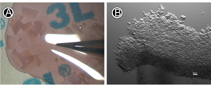
Figure 1: The LNCs isolation process. (A) Corneoscleral rim tissue. (B) Clusters after collagenase A digestion at 37 °C for 18 h. Please click here to view a larger version of this figure.
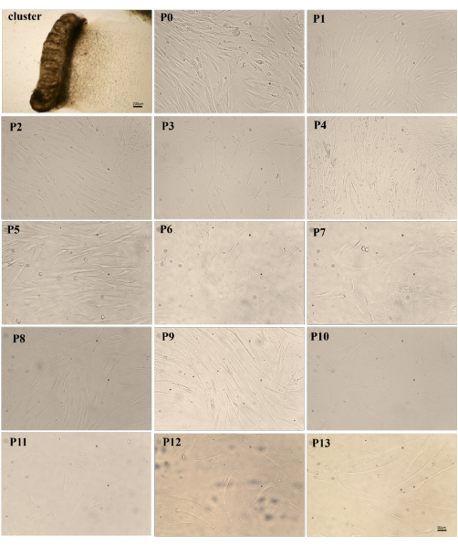
Figure 2: P0 to P13 LNCs cultured on 5% basement membrane matrix coated 6-well plate in MESCM. (Bar = 50 µm). Please click here to view a larger version of this figure.
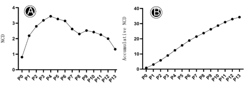
Figure 3: The growth pattern of LNCs from P0-P13. (A) The NCD of LNCs from P0-P13; (B) The cumulative NCD of LNCs from P0-P13. Please click here to view a larger version of this figure.
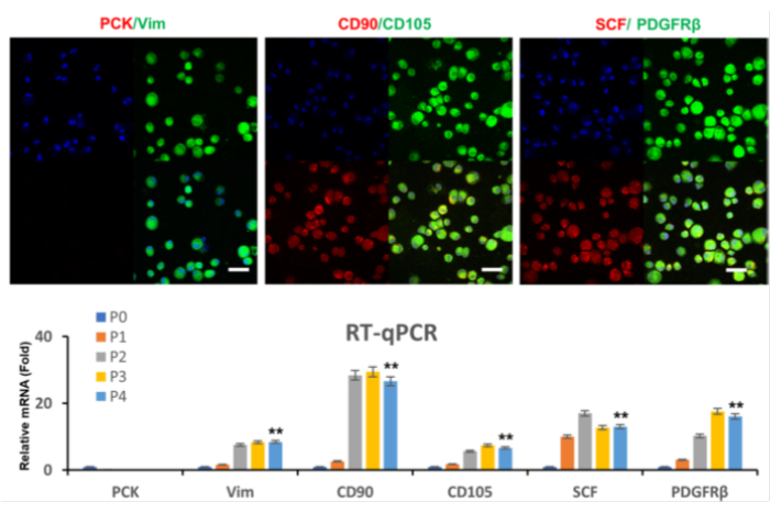
Figure 4: Immunofluorescence and qPCR. Immunofluorescence and qPCR revealed that LNCs uniformly expressed Vim, CD90, CD105, SCF, and PDGFRβ, but not Pan-CK. This figure has been reproduced with permission from Zhu et al.9. Please click here to view a larger version of this figure.
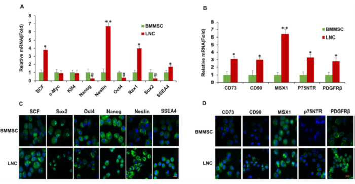
Figure 5: LNCs express more ESC, MSC, and NC markers than BMMSCs. P4 LNCs and P4 BMMSCs were subjected to qPCR for transcription expression of ESC markers (A), MSC and neural crest markers (B) (n = 3, *P < 0.05, #P < 0.05, and **p < 0.01 respectively). Immunostaining of ESC markers (C) and MSC, NC markers (D), with nuclear counterstain by Hoechst 33342. Scale bars = 25 µm. This figure has been reproduced with permission from Li et al.15. Please click here to view a larger version of this figure.
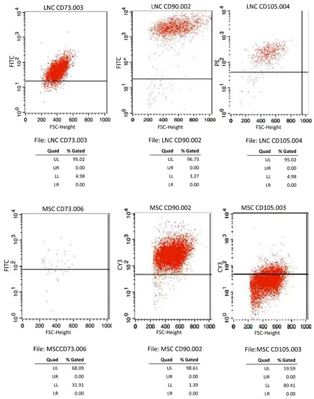
Figure 6: Fluorescence-activated cell sorting of P4 LNCs and BMMSCs (A-F). Fluorescence-activated cell sorting (FACS) analysis of MSC markers including CD73, CD90, and CD105 (n=3). This figure has been reproduced with permission from Li et al.15. Please click here to view a larger version of this figure.
Reagent | Concentration of stock solution | Volume | Final concentration | Storage Environment |
DME/F-12 1:1(1×) | Basic medium | 180 mL | 90% | 4 °C |
KnockOutTMSR | – | 20 mL | 10% | -20 °C |
Serum Replacement for ESCs/iPSCs | ||||
Recominant Human Leukemia Inhibitory Factor (Lif) | 50 µg/mL | 40 µL | 10 ng/mL | -80 °C |
Recombinant Human FGF-basic | 100 µg/mL | 8 µL | 4 ng/mL | -80 °C |
ITS (insulin, transferrin, sodium selenite) | 500 μg/mL insulin | 2 mL | 5 μg/mL insulin | -20 °C |
500 μg/mL transferrin | 5 μg/mL transferrin | |||
500 ng/Ml sodium selenite | 5 ng/mL sodium selenite | |||
Gentamicin | 25 µg/mL | 2 mL | 50 µg/mL | 4 °C |
Amphotericin B | 2500 µg/mL | 100 µL | 1.25 µg/mL | 4 °C |
Table 1: MESCM formulation.
Passage | Seeding Density (× 105 cells/cm2) | Final density (× 105cells/cm2) | Culture time (days) | Number of Cell Doublings (NCD) | Accumulative NCD |
P0 | 0.22 | 0.385714 | 12 | 0.810029 | 0.810029 |
P1 | 0.08 | 0.365714 | 4 | 2.192645 | 3.002674 |
P2 | 0.051429 | 0.357143 | 3 | 2.795859 | 5.798533 |
P3 | 0.037143 | 0.337143 | 2 | 3.182203 | 8.980737 |
P4 | 0.028571 | 0.311429 | 6 | 3.446256 | 12.42699 |
P5 | 0.04 | 0.385714 | 3 | 3.269461 | 15.69645 |
P6 | 0.054286 | 0.48 | 4 | 3.14439 | 18.84084 |
P7 | 0.057143 | 0.351429 | 3 | 2.620586 | 21.46143 |
P8 | 0.08 | 0.394286 | 3 | 2.30117 | 23.7626 |
P9 | 0.06 | 0.345714 | 6 | 2.526546 | 26.28915 |
P10 | 0.022857 | 0.122857 | 7 | 2.426265 | 28.71541 |
P11 | 0.025714 | 0.122857 | 5 | 2.25634 | 30.97175 |
P12 | 0.028571 | 0.114286 | 5 | 2 | 32.97175 |
P13 | 0.045714 | 0.114286 | 10 | 1.321928 | 34.29368 |
Table 2: Serial Passages of the LNC on plastic.
Corneal transparency is typically maintained by regular arrangement and distribution of small fibers (25-30 nm in diameter) in the corneal stroma, which is crucial for normal vision acuity16. There are 253 million visually impaired people worldwide, 36 million of whom are blind17. The world health organization (WHO) considers corneal blindness one of the most serious hazards to human eyesight, accounting for 5.1% of all blindness worldwide16. Corneal epithelium defect with normal CES can heal quickly without leaving a scar18. It is estimated that more than 12.7 million patients worldwide require corneal transplants due to moderate to severe vision loss19; 30% of the cornea transplantation failure was caused by CESD20. However, because of the shortage of cornea donation in most countries worldwide, only one out of seventy cornea-blind patients could eventually receive corneal transplantation21. Currently, corneal epithelial stem cell transplantation (CEST) can be used to treat CESD22. Patients with monocular CESD can obtain CES from healthy eyes for CEST. However, for patients with bilateral CESD, only allogeneic CES can be obtained for treatment. Immune rejection remains the main reason for the failure of allogeneic CEST. A promising method to reduce the therapeutic treatment for CESD is to find a cell that can effectively replace autogenous CES or encourage the differentiation of other autogenous cells from other places to become CES. In vitro differentiation of autologous cells into CES is also becoming popular, including BMMSC23, oral mucosal epithelial cells (OMEC)24, dental pulp stem cells (DPSC)19, human fibroblast-derived-induced pluripotent stem cells (iPSC)25, and adipose stem cells (ASC)26. Rohaina et al.23found that BMMSCs cultured on amniotic membranes for 10 days could differentiate into corneal epithelial cells and that CK3 and p63 expression increased significantly after the induction of differentiation. In 2020, O'Callaghan et al.24induced the differentiation of oral mucosal epithelial cells into corneal epithelial cells using a new culture system, and successfully used it for treating CESD using a 3D tissue structure (RAFT) as a support for oral mucosal epithelial cell culture with human oral mucosal fibroblasts as trophoblasts. Additionally, DPSC differentiates successfully into corneal stromal and epithelial cells. By upregulating the expression of K3, K12, and CD90, DPSC can prevent corneal conjunctival invasion. Another study used DPSC as amniotic cell sheets piggybacked onto the corneal surface in a rabbit CESD model. The results showed that the DPSC group had cleaner corneas and less angiogenesis than the control group27. Hayashi et al.28achieved differentiation using fibroblast-derived iPSC that could be induced in PAX6(+) and K12(+) corneal epithelial cells after 12 weeks. iPSC effectively develops into corneal epithelial cells, activates K12, and suppresses NANOG25. Zeppieri et al.26 discovered that ASCs could differentiate into corneal epithelial cells to cure mouse CESD and enhance corneal epithelial wound healing in a laser-induced animal model.
LNCs localize in the limbal niche, where CES are protected and supported, and have many characteristics similar to MSCs. LNCs were first isolated by Polisetty et al.2 and first named by Xie et al.3. According to recent studies, LNCs are more fundamental stem cells than MSCs and can easily differentiate into adipocytes, chondrocytes, and osteocytes4,29. Compared to the commonly used BMMSCs, LNCs show greater stem cell characteristics (higher expression of CD73, CD105, PDGFR, SCF, etc.) (Figure 5)30. LNCs can successfully treat rabbits with CESD caused by corneal alkali burns by promoting the repair of the corneal epithelium and stroma15. The reduced expression of fibronectin (FN), connective tissue growth factor (CTGF), and secreted protein acidic and cysteine rich (SPARC) in corneal fibroblasts suggest LNCs secretion enhances wound healing and reduces fibrosis7.
In a 3D microenvironment, what is interesting is that LNCs may reunite with MCECs, stimulating the latter cells to restore stem cell properties9. LNCs effectively accelerate CES growth31 and prevents corneal scarring10. However, the quantity of LNCs is limited; studies have indicated it represents only approximately 3% of the total number of limbal stromal cells in the bovine cornea32, with significant expression of keratan sulfate, keratocan, and ALDH3A133. They may be less than 1% in human corneas33.
In this protocol, previous methods of isolating, purifying, and identifying LNCs were repeated3,14,34. After 18 h of treatment of human limbus tissue with collagenase A (2 mg/mL), LNCs were isolated. P0 LNC developed slowly and exhibited various cell morphologies, including spindle-shaped LNCs, polygonal others, and visible spherical MCEC (Figure 2, P0). Previous studies have shown that LNCs are spindle-shaped cells that attach to the bottom of the culture surface with homogeneous morphology14. At the same time, MCECs are round29, which is consistent with the morphology of the cells cultured in this protocol. In addition, P0 LNCs grew slowly to the cells growing full approximately 70%-80% of cell density in approximately 12 days. P1-P12 LNC proliferated substantially faster and, in 3-6 days, practically covered the entire culture plate. Following P1, spindle-shaped LNCs adnexal cells significantly outnumbered round MCEC and polygonal cells.
It is clear from the NCD values in Table 2 and Figure 3 that the LNCs P0 had the lowest NCD (0.810) and that cell passage occurred over 12 days. However, after the P1, the growth of LNCs accelerated dramatically, and the growth rate peaked at 3.44 in the P4. After the P6, the growth rate of the LNCs were substantially reduced (Figure 3). The LNCs growth rate and activity of the P4 were substantially the highest in terms of the LNCs growth pattern, consistent with earlier findings indicating that the P4-P6 LNCs growth activity is the best9. However, this method has certain limitations, the number of LNC P0 varies greatly according to the donor's age and the time of death. Therefore, when the donor tissue is younger and fresher, more P0 LNC can be isolated. The most important factor in mimicking the microenvironment of CES in vitro is the need for universal niche factors, such as a laminamine-rich extracellular matrix35.
In conclusion, LNCs play a crucial role in supporting corneal epithelial stem cells and maintaining the stemness of CES, including self-renewal and differentiation into mature cornea epithelial cells. LNC could be expected to be an innovative cell tool for treating patients with CESD9,15.
The authors have nothing to disclose.
Thanks to Wei Wang, Lingjuan Xu, and Rong Liu for the guidance on this work, Yongyao Tan, Bihui Jin, Chunxiu You, and Li Guigang for providing some of the material, Guanyu Su for writing the manuscript, Xiao Zhou, Yihong Xiong, and Huatao Xie for correcting the manuscript, and Guigang Li for his full guidance. This study was supported by the National Natural Science Foundation of China (No. 82070936, 81470606, 81570819), Hubei Province health and family planning scientific research project (No. WJ2017M073), Top Ten Translational Medical Research Projects from Tongji Hospital (No.2016ZHYX20), Training Project of Young medical Pioneers in Wuhan City (No.2015whzqnyxggrc10), Global Talents Recruitment Program (G2022154028L), National Health Commission of Hubei Province project In 2022(WJ2021ZH0005), and Subject Construction Foundation of Finance Department of Hubei In 2022(42000022815T000000102)
4',6-Diamidino-2-Phenylindole | ThermoFisher | D1306 | 5μg/mL |
Amphotericin B | Sigma | V900919 | 1.25 μg/mL |
Anti-CD73 | Abcam | ab202122 | 1:50 |
Bovine Serum Albumin | MERCK | A1933 | – |
CD105 | Proteintech | 67075-1-Ig | 1:200 |
CD105 | Abcam | ab114052 | 1:50 |
CD90 | Proteintech | 66766-1-Ig | 1:100 |
CD90 | Abcam | ab307736 | 1:50 |
Cell Incubator | Shanghai Lishen | K1119K4644 | HF90(HT) |
Centrifuge system | StatSpin | StatSpin CytoFuge 12 | – |
Collagenase A | Roche | 10103578001 | 2 mg/mL |
Confocal microscope | Zeiss | LSM700 | – |
Culture plate | virya | 3500356 | 35 mm |
DME/F-12 1:1 (1x) | cytiva | SH30023.01 | 90% |
Donkey anti-Mouse IgG (H+L) Secondary Antibody | ThermoFisher | A16016 | 1:1000 |
Donkey anti-rabbit IgG (H+L) Secondary Antibody | ThermoFisher | 31568 | 1:1000 |
FACS Diva sofware | BD Biosciences | Tree Star | – |
Flow Cytometer | BD Biosciences | Becton Dickinson LSRII | – |
Fluorescence microscope | olympus | cx31 | |
Gentamicin | Sigma | G1914 | 50 μg/mL |
Hemocytometer | MERCK | Z359629 | Bright-Line |
High-capacity cDNA Transcription Kit | ThermoFisher | 4374966 | |
Inverted phase-contrast microscope | UOP | DSZ2000X | |
ITS (insulin, transferrin, sodium selenite) | Sigma | I3146 | 5 μg/mL insulin, 5 μg/mL transferrin, 5 ng/mL sodium selenite |
KnockOut SR Serum Replacement for ESCs/iPSCs | gibco | 10828-028 | 10% |
Matrigel | BioCoat | 356234 | – |
Pan-CK | Abcam | ab7753 | 1:1000 |
Paraformaldehyde | NoninBio | NBS0135 | 4.00% |
Paraformaldehyde | MKBio | MM-1505 | 4% |
PDGFRβ | Abclonal | A1444 | 1:100 |
Real-time fluorescence quantitative PCR instrument | Applied Biosystems | Step One Plus | – |
Recombinant Human FGF-basic | Peprotech | 100-18B | 4 ng/mL |
Recominant Human Leukemia Inhibitory Factor(Lif) | Peprotech | 300-05 | 10 ng/mL |
RNeasy Mini RNA Isolation Kit | Qiagen | 74104 | – |
SCF | Bioss | bs-0545R | 1:100 |
SCF | Abcam | ab52603 | 1:50 |
Stereomicroscope | ZEISS | SteREO Discovery. V8 | |
Sterile surgical round blade | Careforde | 29500 | size 10 |
TaqMan Gene Expression Assay Mix | Applied Biosystems | 4448489 | |
Triton X-100 | MERCK | X100 | 0.20% |
Trypan blue | ThermoFisher | 15250061 | 0.40% |
Trypsin-EDTA | Genview | GP3108 | 0.25% |
Tween 20 | MERCK | P9416 | – |
Ultra Clean Bench | LaiTe | LT20200705 | SW-CJ-IFDG |
Universal PCR Master Mix | Applied Biosystems | 4304437 | |
Vim | Abcam | ab92547 | 1:100 |