Summary

基于化学发光的检测方法,用于检测自氧化和亚硝化化合物中的一氧化氮及其衍生物

Published: February 16, 2022
doi:

Summary

在这里,我们提出了使用基于化学发光的高灵敏度测定来检测一氧化氮及其生物相关衍生物的方案。

Abstract

体内一氧化氮(NO)活性是其直接作用,其衍生物由NO自氧化产生的作用以及亚硝化化合物的作用的综合结果。测量NO代谢物对于研究血管水平和其他组织中的NO活性至关重要,特别是在施用外源性NO的实验环境中。基于臭氧的化学发光测定可以精确测量液体(包括血浆,组织匀浆,细胞培养物)和气体混合物(例如,呼出的呼吸)中的NO和NO代谢物。NO与臭氧反应,在激发态下生成二氧化氮。随之而来的光发射允许光探测和产生反映样品NO含量的电信号。来自同一样品的等分试样可用于测量特定的NO代谢物,例如硝酸盐,亚硝酸盐,S-亚硝基硫醇和铁 – 亚硝酰基复合物。此外,无细胞血红蛋白消耗的NO也通过化学发光分析进行量化。提供了所有这些技术的说明。

Introduction

自从萨尔瓦多·蒙卡达(Salvador Moncada)和诺贝尔奖获得者罗伯特·富奇戈特(Robert Furchgott)、路易斯·伊格纳罗(Louis Ignarro)和费里德·穆拉德(Ferid Murad)将一氧化氮(NO)确定为先前已知的内皮源性松弛因子(EDRF)以来,NO的核心作用已在整个血管生物学、神经科学、代谢和宿主反应1234567等几个关键机制中得到确立。.外源性给予NO气体已成为新生儿肺动脉高压引起的呼吸衰竭的既定治疗方法8。一氧化氮气体也已被研究用于治疗呼吸道合胞病毒(RSV)感染,疟疾和其他感染性疾病,缺血再灌注损伤以及预防接受心脏手术的患者的急性肾损伤9101112。对精确测量技术的需求,以评估NO,其代谢物及其靶蛋白和化合物的水平来自机制和介入研究。

由于其高反应性,NO可能会根据其产生和/或释放的生物基质进行不同的反应。在没有血红蛋白(Hb)或其他氧血蛋白的情况下,NO几乎完全氧化成亚硝酸盐(NO2)。

2常开 + O2 → 2常开 2

2 + 否 → N2O3

N2O3 + H2O → NO2 + H+

NO首先与分子氧(O2)进行自氧化以产生二氧化氮(NO2),并与NO2 本身反应生成三氧化二氮(N2O3)。一个分子N2O3 与水(H2O)反应形成两个NO2-分子和 一个质子(H+13。在全血中,1415,NO和NO2 迅速转化为硝酸盐(NO3),因为这些分子与Hb的氧化血红素基团[Hb-Fe2 + -O2 或氧合血红蛋白(oxyHb)]发生激烈反应以产生NO3。该反应与血红素基团向铁态[Hb-Fe3 + 或高铁血红蛋白(metHb)]的转变相结合:

Hb-Fe2+-O2 + NO → Hb-Fe3+ + NO3

红细胞(RBC)屏障和紧邻内皮的空间是限制这种反应的主要因素,并允许内皮释放的一小部分NO作为EDRF1617。事实上,循环中的无细胞Hb已知会破坏实验和临床环境中的血管舒张1718。在红细胞内,根据氧合和NO2 浓度,一部分NO与脱氧血红蛋白(Hb-Fe2 +)反应形成亚硝基铁Hb(Hb-Fe2 + – NO或HbNO):

Hb-Fe2+ + NO → Hb-Fe2+-NO

在RBC1517中,NO2 可以通过减少Hb-Fe 2+ 形成Hb-Fe3+ ,导致NO的释放,而NO又结合Hb-Fe2+-O2 (优先)或Hb-Fe2+

NO衍生物的产生不应被视为严格单向的,因为NO可以从各种组织和不同酶(例如,肠道细菌或线粒体内,特别是在缺氧条件下)的NO2和NO3再生1920

产生(或施用)的可变量的NO导致S-亚硝基硫醇的下游生成,主要是通过在亲核试剂存在下从N2O3 中转硝化硫醇,产生NO + 供体中间体(Nuc-NO+-NO2):

N2O3 + RS → RS-NO + NO2

S-亚硝基硫醇产生的另一种可能性是氧化硫醇的亚硝基化(不与氧化硫醇反应):

RS• + NO → RS-NO

这种机理和NO2 的直接硫醇氧化可能只有在非常具体的条件下才有可能,这些条件在别处21中进行了描述。S-亚硝基硫醇的范围从轻分子(如S-亚硝基谷胱甘肽)到大的含硫醇蛋白质。S-亚硝基血红蛋白(S-NO-Hb)是通过β链(β93C)22中保守半胱氨酸残基的硫醇基团亚硝化形成的。

S-亚硝基硫醇的产生和代谢是重要调节机制的一部分。例子包括调节谷胱甘肽,半胱天冬酶,N-甲基-D-天冬氨酸(NMDA)和ryanodine受体232425262728。亚硝化白蛋白(S-亚硝基白蛋白)先前被认为是体内NO生物学的主要介质,似乎是一种NO / NO + 转运蛋白,没有任何特定的附加生物活性29

在测量生物基质中特定生物样品的NO及其衍生物的浓度时,重要的是要考虑酸度,氧化,温度和试剂的存在等特性。例子包括施用外源性NO供体,并且在急性炎症的情况下,过氧化氢(H2O2)与NO2 反应导致产生过氧亚硝酸盐(ONOO21等自由基的超正常浓度。除了采用的分析方法外,样品制备和储存的预分析阶段也是基础。应预测、考虑和阻断不代表 体内 NO活性的下游反应。一个有效的例子是S-NO-Hb的不稳定性,当血液样本被靶向测量22时,需要对血液样本进行专门的处理。

基于化学发光的测定是检测任何生物体液中NO及其主要代谢物[NO2,NO3-,S-NO和铁- 亚硝酰基复合物(Fe-NO)]水平的金标准,包括组织匀浆3031。这些方法依赖于化学发光检测器(CLD),这是一种容纳NO与臭氧(O3)反应的装置,在激发态(NO2 •)下产生NO2。NO2•的弛豫引起光子的发射,光子由光电倍增管检测到,产生与采样气体混合物32的NO含量成正比的电信号。表示CLD的简化原理图。

Figure 1
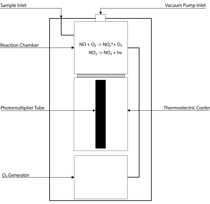
图 1:一氧化氮气体化学发光检测器的简化示意图。 基于化学发光的一氧化氮(NO)检测是在化学发光检测器(CLD)中引入的每个NO气体分子的化学计量生成一个光子。化学发光反应是在指定的腔室中获得的,该腔室由内部发生器提供臭氧(O3),该发生器通过与外部泵连接保持在负压,允许样品气体连续和恒定地流入。O3 的产生需要双原子氧(O2),该氧气由与CLD连接的专用O2 罐供应(其他制造商提供使用环境空气操作的CLD)。在反应室内,样品气体中含有的每一分子NO气体与氧气反应,产生一分子处于活化状态(NO2 *)的二氧化氮。通过返回到其基态,每个NO2 *分子发射一个光子,该光子由位于反应室附近的光电倍增管(PMT)检测。PMT与相关的放大器和中央处理单元产生与光子数和反应室中NO分子数量成比例的信号。 请点击此处查看此图的大图。

CLD的进样口可以连接到包含液体样品反应室的玻璃器皿系统。系统用氮气、氦气或氩气等惰性气体连续吹扫,将NO从反应室转移到CLD。液相样品通过专用膜注入吹扫容器。

Figure 2
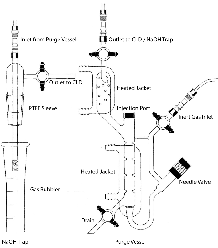
图 2:用于化学发光检测一氧化氮气体的吹扫容器的结构 吹扫容器(右)允许检测一氧化氮 (NO) 气体或任何其他化合物,当从液相试剂中释放时,可以很容易地转化为 NO 气体。惰性气体入口连接到惰性气体的源(罐),例如氩气,至强或双原子氮(N2)。针阀(向左打开)用于吹扫容器内的压力控制,可以完全拆卸以清洁容器。进样口由带有膜隔的盖子覆盖,用于样品进样。应经常更换膜。加热的夹套包围反应室并连接到热水浴中以在HCl测定中进行VCl3 。吹扫容器出口连接到化学发光检测器(CLD)或NaOH捕集器(HCl测定中VCl3 需要)。要排出反应室内容物,首先关闭惰性气体入口和吹扫容器出口处的旋塞阀,关闭针阀,取下注射口处的盖子,最后打开排放口处的旋塞阀。如果由于HCl的腐蚀性而进行HCl测定中的VCl3 ,则需要将NaOH陷阱(左)放置在吹扫容器和CLD之间。与CLD的连接始终需要在CLD和吹扫容器(或NaOH陷阱,如果使用)的输出之间放置一个强场电介质(IFD)滤波器。IFD过滤器去除空气中的颗粒物,并阻止液体通过吹扫容器。PTFE = 聚四氟乙烯。 请点击此处查看此图的大图。

因此,任何可以通过特定和受控化学反应转化为NO的化合物都可以在任何生物体液和组织匀浆24中以高灵敏度检测到。通过化学发光直接测量气相NO是在实验和临床环境中进行的。这些技术在别处有333435的广泛描述。NO2、S-亚硝基硫醇、S-亚硝化物蛋白和Fe-NOs的测量可以通过在三碘化物(I 3-)的反应混合物中加入样品来进行,三碘化物(I3)化学计量法从所有这些化合物中释放出NO气体:

I3 → I2 + I

2常2 +2I +4H+ → 2常数 + I2 +2H2O

I3 + 2RS-NO → 3I + RSSR + 2NO+

2NO+ + 2I → 2NO + I2

而I3不与NO 3-15反应。通过用酸化磺胺(AS)预处理样品等分试样(含或不含氯化汞(HgCl2),可以对每种化合物进行精确测量。具体而言,AS预处理去除所有NO2含量。因此,CLD测量的NO含量仅反映了S-NOs和Fe-NOs浓度的总和。在AS注射之前,在样品等分试样中注射HgCl 2会导致NO2由S-NO释放。用AS处理(导致NO2去除)确保测量的NO含量仅反映Fe-NOs的浓度。评估之间的一系列减法允许计算三个NO导数22的精确浓度。
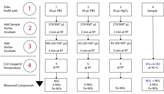
Figure 3
图3:在乙酸化学发光测定中I3样品制备的步骤。说明了制备乙酸化学发光测定中I3的顺序步骤。需要使用避光离心管。试管1,2和3是用于准备测定的试管。如果需要测量硝酸盐(NO3),则需要另一个样品等分试样(管4)用于HCl测定中的VCl3。步骤由红色的数字表示。在加入样品体积之前,用磷酸盐缓冲盐水(PBS)或HgCl2预填充(步骤1)。按照指示加入样品体积(2),涡旋,并在室温(RT)下孵育2分钟。按照指示加入(3)PBS或酸化磺胺(AS),涡旋,并在室温下孵育3分钟。通过测定测量的浓度是每个管下报告的化合物浓度的总和。1号管将允许测量亚硝酸盐(NO2),S-亚硝基硫醇(S-NO)和铁 – 亚硝基配合物(Fe-NOs)作为单个信号。对于硝酸盐(NO3)测量,样品应同时在乙酸中与I3和VCl3一起在HCl测定中运行,并且从管1获得的值应从从管4中获得的值中减去。*建议用于Hb分析以测定残留NO2,S-亚硝基血红蛋白和铁-亚硝酰血红蛋白的量。请点击此处查看此图的大图。

对于 NO3 测量,盐酸 (HCl) 中的氯化钒 (III) (VCl3) 用于将吹扫容器中的 NO3 转化为 NO,以便使用 CLD 以化学计量法测量 NO3

2 无3+ 3V+3 + 2H2O → 2NO + 3VO2+ + 4H+

为了实现足够快的转化,反应需要在90-95°C下进行。 从NO3 减少到NO2 与HCl将NO2 还原为NO相结合,钒金属也减少了S-NOs,释放了它们的NO部分2236。CLD在HCl中用VCl3 获得的最终浓度反映了NO3,NO2和其他亚硝化化合物的总浓度。用I3 从CLD产生的浓度中减去后一个值,可以计算NO3 浓度3637图3)。

在NO消耗测定中,NO供体如(Z)-1-[2-(2-氨基乙基)-N-(2-氨乙基)氨基]二禅-1-鎓-1,2-二乙酸(DETA-NONO酸酯)在吹扫容器中连续释放NO产生稳定的信号,允许定量注射样品中的无细胞氧Hb。吹扫容器中消耗的NO的量与样品38中oxyHb的量呈化学计量关系。

图中说明了测量血浆样品中 NO2、NO3-、S-亚硝基硫醇、亚硝酰铁配合物和无细胞 Hb 消耗的实验方案。对红细胞环境中的NO的研究需要特定的样品处理,然后进行排阻色谱法以测量极其脆弱的S-NO-Hb和Hb-NO,并测定总Hb浓度1522。样品制备有助于校正测量。在测定过程中,H2 O中NO2的预先存在和NO2的释放可导致人为测量更高浓度的NO衍生物,例如S-NO-Hb1439。还介绍了样品制备的重要方面。

Protocol

该协议中指明的程序符合马萨诸塞州总医院的审查委员会。所使用的血液样本是在先前的一项研究中收集的,并且出于当前目的进行了去识别化 18. 注:有关管道和构成吹扫容器的玻璃器皿之间的最佳连接、清洗和一般维护的具体指导,请参阅制造商的说明。连接需要牢固且小心,以免损坏玻璃器皿。识别玻璃吹扫容器的组件:进气管路、带加热夹套和冷凝?…

Representative Results

通过无细胞Hb测定的NO消耗用于含有已知浓度的无细胞氧Hb的样品(图4)。由于氧肝素的一个血红素在化学计量法中释放出一个NO分子,因此纯化的无细胞氧肝素用于构建测定的校准曲线(补充图3)。 无细胞Hb(用比色测定法测量)与心脏手术期间体外循环(CPB)患者NO消耗之间的剂量 – 反应关系如图 5所示。可以假设?…

Discussion

由于灵敏度高,用于测定NO和相关化合物的基于化学发光的测定具有NO2 污染的高风险。实验中使用的每种试剂(特别是NO2 封闭溶液)和稀释剂(包括ddH2O)都应测试其NO2 含量以校正背景信号。亚硝酸盐反应性极强,全血半衰期约为10分钟,并迅速产生NO3。因此,血液采集和离心之间的时间或NO2 阻断可能…

Disclosures

The authors have nothing to disclose.

Acknowledgements

本手稿中报告的协议是由马萨诸塞州总医院麻醉科Warren Zapol博士的重症监护麻醉研究实验室的先前研究员的累积贡献而实现的。我们感谢Akito Nakagawa,Francesco Zadek,Emanuele Vassena,Chong Lei,Yasuko Nagasaka,Ester Spagnolli和Emanuele Rezoagli博士的贡献。

Materials

Acetic Acid Sigma 45754 500 mL – liquid
Antifoam B Emulsion Sigma A5757 250 mL – liquid
DETA NONOate Cayman 82120 10 mg
Gibco DPBS (1x) no calcium, no magnesium ThermoFisher 14190144 500 mL
Hydroochloric Acid 37% (1 M) Sigma 258148 500 mL – liquid
Iodine SAFC 207772 100 g – solid
Kimwipes Kimtech 34155
Mercury (II) Chloride ACS reagent> 99.5% Sigma 215465 100 g – solid (dissolve in water)
Mili-Q Water Purification System Millipore
Model 705 RN 50 μL syringe Hamilton 80530 Microliter syringe
Model 802 N 25 μL Syringe Hamilton 84854 Microliter syringe
N-ethylmaleimide Sigma 4260 25 g – crystalline
Nitric Oxide Analyzer + Bundle Software – Purge Vessel Zysense NOA 280i Chemiluminescence Detector
Nonidet p-40  (NP-40) ThermoFisher 85125 10% – 500 mL
Potassium hexacyanoferrate (III) ACS reagent≥ 99% Sigma 244023 100 g – powder
Potassium Iodide ACS reagent> 99% Sigma 221945 100 g – solid
Potassium Nitrite cryst. For analysis EMSURE ACS Supelco 105067 250 g – crystalline
PowerGen 125 Fisher Scientific 14-359-251 Mechanic Homogenizer
RV3 Two Stage Rotary Vane Pump Edwards A65201906 Vacuum Pump – Bundled with analyzer
Sodium Heparin – BD Hemogard Clo BD Biosciences BD367871 75 USP Units
Sodium hydroxide anhydrous ACS reagent ≥ 97% Sigma 795429 1 kg – pelletts
Sodium Nitrate ACS reagent ≥ 99% Sigma 221341 500 g – powder
Sodium nitrite ≥ 99% Sigma S2252 500 g – crystalline
Sulfanilamide ≥ 98% Sigma S9251 100 g – solid
Vanadium (III) Chloride Sigma 112393 25 g – solid – Caution (exothermic)
Whatman 1 Filter Paper Sigma WHA10010155

References

  1. Palmer, R. M. J., Ferrige, A. G., Moncada, S. Nitric oxide release accounts for the biological activity of endothelium-derived relaxing factor. Nature. 327 (6122), 524-526 (1987).
  2. Furchgott, R. F. The discovery of endothelium-derived relaxing factor and its importance in the identification of nitric oxide. JAMA: The Journal of the American Medical Association. 276 (14), 1186 (1996).
  3. Ignarro, L. J., Buga, G. M., Byrns, R. E., Wood, K. S., Chaudhuri, G. Endothelium-derived relaxing factor and nitric oxide possess identical pharmacologic properties as relaxants of bovine arterial and venous smooth muscle. The Journal of Pharmacology and Experimental Therapeutics. 246 (1), 218-226 (1998).
  4. Arnold, W. P., Mittal, C. K., Katsuki, S., Murad, F. Nitric oxide activates guanylate cyclase and increases guanosine 3′:5′-cyclic monophosphate levels in various tissue preparations. Proceedings of the National Academy of Sciences of the United States of America. 74 (8), 3203-3207 (1977).
  5. Hayashida, K., et al. Depletion of vascular nitric oxide contributes to poor outcomes after cardiac arrest. American Journal of Respiratory and Critical Care Medicine. 199 (10), 1288-1290 (2019).
  6. Ignarro, L. J. Inhaled NO and COVID-19. British Journal of Pharmacology. 177 (16), 3848-3849 (2020).
  7. Gantner, B. N., LaFond, K. M., Bonini, M. G. Nitric oxide in cellular adaptation and disease. Redox Biology. 34, 101550 (2020).
  8. Clark, R. H., et al. Low-dose nitric oxide therapy for persistent pulmonary hypertension of the newborn. New England Journal of Medicine. 342 (7), 469-474 (2000).
  9. Goldbart, A., et al. Inhaled nitric oxide therapy in acute bronchiolitis: A multicenter randomized clinical trial. Scientific Reports. 10 (1), (2020).
  10. Bangirana, P., et al. Inhaled nitric oxide and cognition in pediatric severe malaria: A randomized double-blind placebo controlled trial. PloS One. 13 (1), 0191550 (2018).
  11. Jiang, S., Dandu, C., Geng, X. Clinical application of nitric oxide in ischemia and reperfusion injury: A literature review. Brain Circulation. 6 (4), 248-253 (2020).
  12. Lei, C., et al. Nitric oxide decreases acute kidney injury and stage 3 chronic kidney disease after cardiac surgery. American Journal of Respiratory and Critical Care Medicine. 198 (10), 1279-1287 (2018).
  13. Kelm, M. Nitric oxide metabolism and breakdown. Biochimica et Biophysica Acta. 1411 (2-3), 273-289 (1999).
  14. Bryan, N. S., Grisham, M. B. Methods to detect nitric oxide and its metabolites in biological samples. Free Radical Biology and Medicine. 43 (5), 645-657 (2007).
  15. Macarthur, P. H., Shiva, S., Gladwin, M. T. Measurement of circulating nitrite and S-nitrosothiols by reductive chemiluminescence. Journal of Chromatography B. 851 (1-2), 93-105 (2007).
  16. Helms, C., Kim-Shapiro, D. B. Hemoglobin-mediated nitric oxide signaling. Free Radical Biology and Medicine. 61, 464-472 (2013).
  17. Kim-Shapiro, D. B., Schechter, A. N., Gladwin, M. T. Unraveling the reactions of nitric oxide, nitrite, and hemoglobin in physiology and therapeutics. Arteriosclerosis, Thrombosis, and Vascular Biology. 26 (4), 697-705 (2006).
  18. Rezoagli, E., et al. Pulmonary and systemic vascular resistances after cardiopulmonary bypass: role of hemolysis. Journal of Cardiothoracic and Vascular Anesthesia. 31 (2), 505-515 (2017).
  19. Shiva, S. Nitrite: A physiological store of nitric oxide and modulator of mitochondrial function. Redox Biology. 1 (1), 40-44 (2013).
  20. Lundberg, J. O., Weitzberg, E., Gladwin, M. T. The nitrate-nitrite-nitric oxide pathway in physiology and therapeutics. Nature Reviews Drug Discovery. 7 (2), 156-167 (2008).
  21. Heinrich, T. A., Da Silva, R. S., Miranda, K. M., Switzer, C. H., Wink, D. A., Fukuto, J. M. Biological nitric oxide signalling: chemistry and terminology. British Journal of Pharmacology. 169 (7), 1417-1429 (2013).
  22. Yang, B. K., Vivas, E. X., Reiter, C. D., Gladwin, M. T. Methodologies for the sensitive and specific measurement of s -nitrosothiols, iron-nitrosyls, and nitrite in biological samples. Free Radical Research. 37 (1), 1-10 (2003).
  23. Hayashida, K., et al. Improvement in outcomes after cardiac arrest and resuscitation by inhibition of s-nitrosoglutathione reductase. Circulation. 139 (6), 815-827 (2019).
  24. Rodríguez-Ortigosa, C. M., et al. Biliary secretion of S-nitrosoglutathione is involved in the hypercholeresis induced by ursodeoxycholic acid in the normal rat. Hepatology. 52 (2), 667-677 (2010).
  25. Mitchell, D. A., Marletta, M. A. Thioredoxin catalyzes the S-nitrosation of the caspase-3 active site cysteine. Nature Chemical Biology. 1 (3), 154-158 (2005).
  26. Mannick, J. B., et al. Fas-induced caspase denitrosylation. Science. 284 (5414), 651-654 (1999).
  27. Choi, Y. -. B., et al. Molecular basis of NMDA receptor-coupled ion channel modulation by S-nitrosylation. Nature Neuroscience. 3 (1), 15-21 (2000).
  28. Eu, J. P., Sun, J., Xu, L., Stamler, J. S., Meissner, G. The skeletal muscle calcium release channel: Coupled O2 sensor and NO signaling functions. Cell. 102 (4), 499-509 (2000).
  29. Stamler, J. S., et al. Nitric oxide circulates in mammalian plasma primarily as an S-nitroso adduct of serum albumin. Proceedings of the National Academy of Sciences of the United States of America. 89 (16), 7674-7677 (1992).
  30. Dunham, A. J., Barkley, R. M., Sievers, R. E. Aqueous nitrite ion determination by selective reduction and gas phase nitric oxide chemiluminescence. Analytical Chemistry. 67 (1), 220-224 (1995).
  31. Hogg, N., Zielonka, J., Kalyanaraman, B., Ignarro, L. J., Freeman, B. A. Detection of Nitric Oxide and Peroxynitrite in Biological Systems: A State-of-the-Art Review. Nitric Oxide (Third Edition). , 23-44 (2017).
  32. Gudem, M., Hazra, A. Mechanism of the chemiluminescent reaction between nitric oxide and ozone. The Journal of Physical Chemistry A. 123 (4), 715-722 (2019).
  33. Ishibe, Y., Liu, R., Hirosawa, J., Kawamura, K., Yamasaki, K., Saito, N. Exhaled nitric oxide level decreases after cardiopulmonary bypass in adult patients. Critical Care Medicine. 28 (12), 3823-3827 (2000).
  34. American Thoracic Society, European Respiratory Society. ATS/ERS recommendations for standardized procedures for the online and offline measurement of exhaled lower respiratory nitric oxide and nasal nitric oxide, 2005. American Journal of Respiratory and Critical Care Medicine. 171 (8), 912-930 (2005).
  35. Cuthbertson, B. H., Stott, S. A., Webster, N. R. Exhaled nitric oxide as a marker of lung injury in coronary artery bypass surgery. British Journal of Anaesthesia. 89 (2), 247-250 (2002).
  36. Ewing, J. F., Janero, D. R. Specific S-nitrosothiol (thionitrite) quantification as solution nitrite after vanadium(III) reduction and ozone-chemiluminescent detection. Free Radical Biology and Medicine. 25 (4-5), 621-628 (1998).
  37. Braman, R. S., Hendrix, S. A. Nanogram nitrite and nitrate determination in environmental and biological materials by vanadium(III) reduction with chemiluminescence detection. Analytical Chemistry. 61 (24), 2715-2718 (1989).
  38. Wang, X., et al. Biological activity of nitric oxide in the plasmatic compartment. Proceedings of the National Academy of Sciences of the United States of America. 101 (31), 11477-11482 (2004).
  39. Bryan, N. S., Rassaf, T., Rodriguez, J., Feelisch, M. Bound NO in human red blood cells: fact or artifact. Nitric Oxide. 10 (4), 221-228 (2004).
  40. Kida, K., Shirozu, K., Yu, B., Mandeville, J. B., Bloch, K. D., Ichinose, F. Beneficial effects of nitric oxide on outcomes after cardiac arrest and cardiopulmonary resuscitation in hypothermia-treated mice. Anesthesiology. 120 (4), 880-889 (2014).
  41. Gladwin, M. T., et al. S-Nitrosohemoglobin is unstable in the reductive erythrocyte environment and lacks O2/NO-linked allosteric function. The Journal of Biological Chemistry. 277 (31), 27818-27828 (2002).
  42. Feelisch, M., et al. and heme-nitros(yl)ation in biological tissues and fluids: implications for the fate of NO in vivo. The FASEB Journal. 16 (13), 1775-1785 (2002).
  43. Liu, T., et al. L-NAME releases nitric oxide and potentiates subsequent nitroglycerin-mediated vasodilation. Redox Biology. 26, 101238 (2019).
  44. Piknova, B., Park, J. W., Cassel, K. S., Gilliard, C. N., Schechter, A. N. Measuring nitrite and nitrate, metabolites in the nitric oxide pathway, in biological materials using the chemiluminescence method. Journal of Visualized Experiments: JoVE. (118), e54879 (2016).
  45. Keefer, L. K., Nims, R. W., Davies, K. M., Wink, D. A. 34;NONOates" (1-substituted diazen-1-ium-1,2-diolates) as nitric oxide donors: Convenient nitric oxide dosage forms. Methods in Enzymology. , 281-293 (1996).
  46. Nagababu, E., Rifkind, J. M. Measurement of plasma nitrite by chemiluminescence without interference of S-, N-nitroso and nitrated species. Free Radical Biology and Medicine. 42 (8), 1146-1154 (2007).
  47. Doctor, A., et al. Hemoglobin conformation couples erythrocyte S-nitrosothiol content to O2 gradients. Proceedings of the National Academy of Sciences of the United States of America. 102 (16), 5709-5714 (2005).
  48. Zhang, Y., Keszler, A., Broniowska, K. A., Hogg, N. Characterization and application of the biotin-switch assay for the identification of S-nitrosated proteins. Free Radical Biology and Medicine. 38 (7), 874-881 (2005).
  49. Davies, I. R., Zhang, X., Poole, R. K. Nitric Oxide Selective Electrodes. Methods in enzymology. , 63-95 (2008).
  50. Saito, J., et al. Comparison of fractional exhaled nitric oxide levels measured by different analyzers produced by different manufacturers. Journal of Asthma. 57 (11), 1216-1226 (2020).

Play Video

Cite This Article
Di Fenza, R., Yu, B., Carroll, R. W., Berra, L. Chemiluminescence-based Assays for Detection of Nitric Oxide and its Derivatives from Autoxidation and Nitrosated Compounds. J. Vis. Exp. (180), e63107, doi:10.3791/63107 (2022).

View Video