Summary

発生中の(カエル)胚における細胞系譜ガイド質量分析プロテオミクス

Published: April 21, 2022
doi:

Summary

ここでは、脊椎動物 アフリカツメガエル 胚における既知の組織運命を有する細胞系譜の質量分析ベースのプロテオミクス特性評価について述べる。

Abstract

細胞が組織や臓器を生じさせるときの分子事象の特性評価は、正常な発達をよりよく理解し、病気の効率的な治療法を設計する可能性を高めます。多様な種類と多数のタンパク質の正確な同定と定量を可能にする技術は、空間と時間における組織と生物の発達を調整する分子メカニズムに関するまだ不足している情報を提供します。ここでは、 アフリカツメガ エル(カエル)胚の同定された細胞系譜における数千のタンパク質の測定を可能にする質量分析ベースのプロトコルを紹介します。このアプローチは、再現性のある細胞運命マップと確立された方法に基づいて構築されており、脊椎動物の発生のこのモデルから細胞とその子孫(クローン)を識別、蛍光標識、追跡、およびサンプリングします。マイクロサンプリングまたは解剖または蛍光活性化細胞ソーティングによる細胞単離を使用して細胞内容物を収集した後、タンパク質を抽出し、ボトムアッププロテオミクス解析のために処理します。液体クロマトグラフィーとキャピラリー電気泳動は、高分解能質量分析(HRMS)によるタンパク質の検出と定量のためのスケーラブルな分離を提供するために使用されます。神経組織運命細胞のプロテオミクス特性評価のための代表的な例が提供されています。細胞系譜ガイド下HRMSプロテオミクスは、さまざまな組織や生物に適応できます。脊椎動物の発生中のプロテオームの時空間ダイナミクスを覗き込むのに十分な感度、特異性、定量性を備えています。

Introduction

細胞分化と組織や臓器の起源に関する私たちの理解は、遺伝子とその産物の何十年にもわたる精巧な標的スクリーニングの結果です。重要な細胞イベント中のすべての生体分子とその量に関する知識を増やすことは、脊椎動物のボディプランの空間的および時間的パターンを制御する分子メカニズムを解明するのに役立ちます。分子増幅とシーケンシングを可能にする技術は、現在、多数の遺伝子と転写物について日常的に報告できるようになり、基礎生物学およびトランスレーショナル研究における仮説主導の研究をサポートしています。発達中のシステムを理解するために、転写と翻訳の間の複雑な関係は、複数のタンパク質とその翻訳後修飾の直接分析を提唱しています。人工多能性幹細胞などのin vitro生物学的システムを使用したグローバルプロテオミクスは、組織誘導のメカニズムを描写し始めました1,2。脊椎動物の胚のような複雑な生物では、発生は空間と時間の文脈でモルフォゲン勾配に依存しています3。したがって、細胞が分化して神経組織などの特殊な組織を形成する際のプロテオミクス変化の知識を得ることは、正常および欠陥のある発生を制御する分子プログラムを解き放ち、次世代の治療法を導くための鍵を提供します。

脊椎動物の南アフリカのツメガエル(アフリカツメガエル)は、細胞および発生、神経、および再生生物学において確立されたモデルです。体細胞核の多能性の発見に対するジョン・ガードン卿の2012年ノーベル生理学・医学賞4,5は、基礎研究およびトランスレーショナル研究における発見のためのこのモデルの重要性を強調しました。アフリカツメガエル胚は母親の外部で発生するため、さまざまな発生段階にわたる細胞、細胞クローン、および遺伝子発現の直接操作が容易になります。非対称色素沈着とステレオタイプの細胞分裂により、16-6および32細胞の7,8段階の胚からの再現可能な運命マップのチャート作成が可能になりました。高分解能質量分析(HRMS)ベースのプロテオミクスでは、比較的大きなサイズ(直径~1 mm)で、分析用の豊富なタンパク質含有量が得られる(初期卵割段階の胚では~130 μg、16細胞胚の単一細胞では~10 μgのタンパク質含有量)9,10

現在、HRMSはタンパク質の検出に最適な最先端の技術です。この技術により、複数の、通常は数百から数千の異なるタンパク質の直接的、高感度、特異的な検出と定量が可能になります11。HRMSによるボトムアッププロテオミクスには、相互に関連する一連のステップが含まれます。細胞/組織サンプルからの抽出後、タンパク質はトリプシン(ボトムアッププロテオミクス)などのタンパク質分解酵素で消化されます。得られたペプチドは、疎水性(逆相液体クロマトグラフィー、LC)、正味電荷(イオン交換クロマトグラフィー)、サイズ(サイズ排除クロマトグラフィー)、または電気泳動移動度(キャピラリー電気泳動、CE)など、さまざまな物理化学的特性に基づいて分離されます。次に、通常はエレクトロスプレーイオン化(ESI)を使用してペプチドを荷電(イオン化)し、タンデムHRMSによる気相フラグメンテーションを介してペプチドイオンを検出および配列決定します。得られたペプチドデータは、研究されている生物のプロテオームにマッピングされます。タンパク質特異的(プロテオタイプ)ペプチドイオンシグナル強度は濃度と相関するため、タンパク質定量は、ラベルフリーまたはラベルベース(マルチプレックス定量)で行うことができます。HRMSプロテオミクスは、研究中のシステムの分子状態に関する豊富な情報リソースを生成し、仮説の生成とフォローアップ機能研究を可能にします。

Figure 1
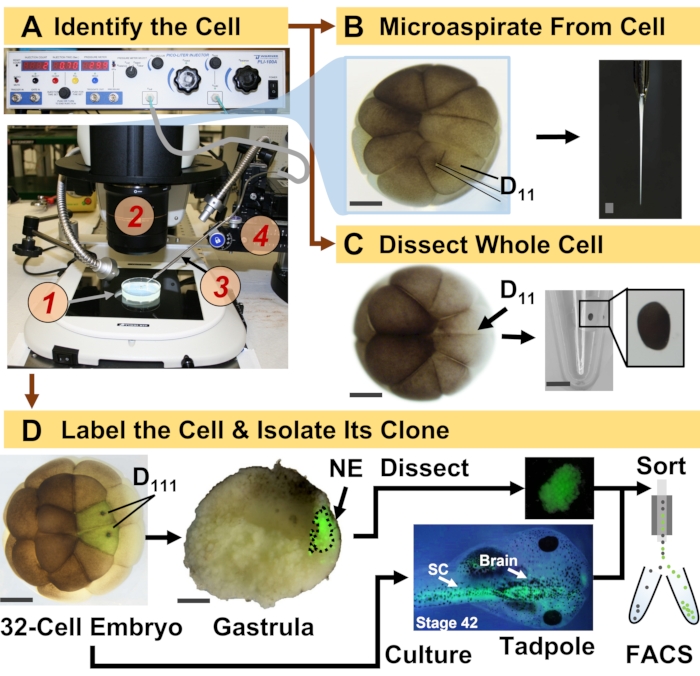
図1:発生中の(カエル)胚における細胞系譜ガイドHRMSプロテオミクスを可能にする時空間的にスケーラブルなプロテオミクス。 (a)標本の可視化(1)を実体顕微鏡を用いて(2)同定した細胞(挿入図)を注入し、作製したマイクロピペット(3)を翻訳段階(4)により制御する。(b)16細胞胚における同定された左D11細胞の細胞内サンプリング。(C)16細胞胚からのD11細胞全体の解剖。(D)原腸内の神経外胚葉(NE)の解剖(ステージ10)をガイドするための、32細胞胚からの左右のD111子孫の蛍光(緑色)追跡、およびFACSを使用したオタマジャクシから子孫組織の分離。スケールバー:胚の場合は200 μm、バイアルの場合は1.25 mm。図は参考文献15,19,21,59の許可を得て翻案した。この図の拡大版を表示するには、ここをクリックしてください。

ここで紹介するプロトコルは、発生中のX. laevis胚で同定された細胞/組織中の多数のタンパク質のHRMSベースの定量を可能にします。このアプローチは、正確な細胞同定、再現性のある細胞運命マップ、およびこの生物学的モデルで細胞系譜を追跡するための確立された方法論に基づいています6,7,8図1に示すように、全細胞解剖またはキャピラリーマイクロサンプリングを使用して細胞含有量を吸引することにより、単一細胞からのプロテオームを研究します。細胞の系統を監視することで、原腸形成中に細胞が組織を形成する際のプロテオームの時空間進化を研究することができます。細胞子孫は、蛍光タンパク質(例えば、緑色蛍光タンパク質、またはGFP)のために不活性デキストランまたはmRNAに結合した蛍光色素を注入することによって蛍光的にマークされる。標識された子孫は、所望の発生時点で単離される。原腸形成中に、密集した細胞クローンは解剖によって単離され得る。原腸形成後、細胞クローンは移動運動のために胚内に分布し、蛍光活性化セルソーティング(FACS)によって解離組織から単離することができます。これらの細胞および組織中のタンパク質は、分離にHPLCまたはCEを使用し、同定にESIタンデムHRMSを使用するボトムアッププロテオミクスを介して測定されます。細胞系譜ガイド下HRMSプロテオミクスは、胚内のさまざまな細胞サイズと系統に拡張可能であり、特異的、高感度、定量的です。ここに示すいくつかの例を通じて、このプロトコルがスケーラブルであり、さまざまな種類の細胞や細胞系譜に広く適応できることも示しています。

Figure 2
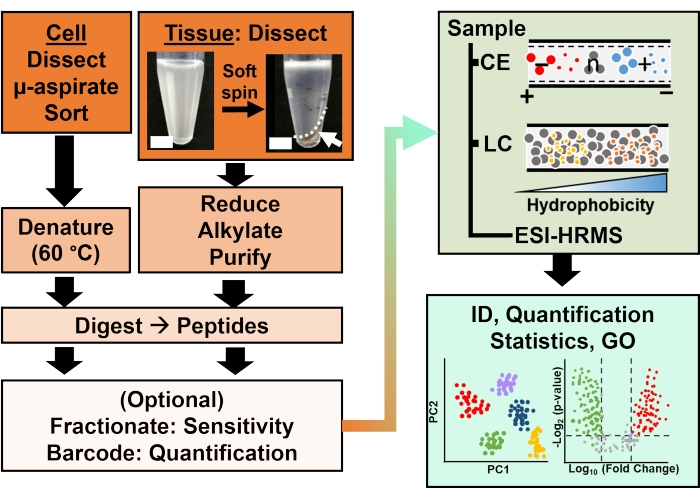
図2:バイオ分析ワークフロー。 微小解剖および毛細血管吸引、またはFACSは、細胞およびクローンタンパク質含有量のサンプリングを容易にしました。豊富な卵黄タンパク質の枯渇とキャピラリー電気泳動(CE)またはナノフロー液体クロマトグラフィー(LC)による分離により、エレクトロスプレーイオン化(ESI)高分解能質量分析(HRMS)を使用した同定(ID)感度が向上しました。定量化により、制御不全が明らかになり、遺伝子オントロジー(GO)から入手可能な情報と組み合わせて、仮説主導の研究に新しい情報を提供しました。図は参考文献15の許可を得て翻案した。 この図の拡大版を表示するには、ここをクリックしてください。

Protocol

アフリカツメガエルの成体のカエルの人道的な維持と取り扱いを保証するすべてのプロトコルは、メリーランド大学カレッジパーク校の施設動物管理および使用委員会によって承認されました(承認番号R-DEC-17-57およびR-FEB-21-07)。 1.ソリューションを準備します 発生学用1x、0.5x、および0.2x Steinberg溶液(SS)を調製し、標準プロトコル<sup class="…

Representative Results

このプロトコルにより、X. laevis胚の組織を確立する際の単一細胞とその系統のタンパク質の研究が可能になりました。図1は、神経組織運命細胞および胚の新たに誘導された神経外胚葉におけるタンパク質を研究するためのアプローチのそのような応用の1つを示しています。図1Aに示すように、バイオアナリシスワークフローは、細胞および…

Discussion

このプロトコルにより、 アフリカツメガエル 種の胚における同定された細胞系譜におけるタンパク質発現の特性評価が可能になります。HRMSに由来するこの方法論は、分子同定における絶妙な特異性、分子プローブ(通常は数百から数千の異なるタンパク質)を使用しないマルチタンパク質検出能力、および定量能力を兼ね備えています。細胞および発生(神経)生物学における古典的なツ…

Disclosures

The authors have nothing to disclose.

Acknowledgements

胚の解離とFACSに関する貴重な議論をしてくれたJie Li(メリーランド大学カレッジパーク校)に感謝します。Vi M. QuachとCamille Lombard-Banekには、このプロトコルで強調されているプロテオミクスアプリケーションを例示する以前の研究におけるサンプル調製とデータ収集を支援していただき、感謝します。この研究の一部は、国立科学財団(賞番号IOS-1832968 CAREER)(P.N.)、国立衛生研究所(賞番号R35GM124755)(P.N.)、メリーランド大学-国立がん研究所パートナーシッププログラム(P.N.)、およびCOSMOSクラブ財団研究賞(A.B.B.およびL.R.P.)によって支援されました。

Materials

Acetonitrile (LC-MS-grade) Fisher Scientific A955
Agarose ThermoFisher Scientific R0492
Ammonium bicarbonate Fisher Scientific A643-500
Analytical Column Thermo Scientific 164941
Analytical microbalance Mettler-Toledo XSE105DU
Automatic peptide fractionation platform Agilent 1260 Infinity II
Borosilicate Capillaries Sutter Instruments Co. B100-50-10
Borosilicate Capillaries (for making Emmitters) Sutter Instruments B100-75-10
C18 spin columns (for desalting) ThermoFisher Scientific 89870
Camera ro monitor electrospray Edmund Optics Inc. EO-2018C
Combretastatin A4 Millipore Sigma C7744
Commercial CESI system AB SCIEX CESI
(Cyclohexylamino)-1-propanesulfonic acid (CAPS) VWR 97061-492
Cytochalasin D Millipore Sigma C8273
Dextran, Alexa Fluor 488; 10,000 MW, Anionic, Fixable ThermoFisher Scientific D22910
Diothiothreitol Fisher Scientific FERR0861
Dumont #5 Forceps Fine Science Tools 11252-30
EDTA Fisher Scientific AAJ62786AP
Epifluorescence light source Lumencore AURA III
Eppendorf LoBing microcentrifuge  tubes: protein Fisher Scientific 13-698-793
Formic acid (LC-MS-grade) Fisher Scientific A117-50
Freezer (-20 °C) Fisher Scientific 97-926-1 
Freezer (-80 °C) Thermo Scientific TSX40086A
Fused silica capillary Molex 1088150596
Heat Block Benchmark BSH300
High pressure liquid Chromatography System ThermoFisher Scientific Dionex Ultimate 3000 RSLC nanosystem
High voltage power supply Spellman CZE1000R
High-resolution Mass Spectrometer ThermoFisher Scientific Orbitrap Fusion Lumos Tribrid Mass Spectrometer
HPLC caps Thermo Scientific C4013-40A
HPLC Vials Thermo Scientific C4013-11
Illuminator e.g. Goosenecks Nikon C-FLED2
Ingenuity Pathway Analysis Qiagen
Iodoacetamide Fisher Scientific AC122275000
Methanol (LC-MS-grade) Fisher Scientific A456
Methanol (LC-MS-grade) Fisher Scientific A456-4
Microcapillary puller Suttor Instruments P-2000
Microinjector Warner Instrument, Handem, CT PLI-100A
Micropippette puller Sutter Instruments Co. P-1000
MS data analysis software, commercial ProteomeDiscoverer
MS data analysis software, opensource MaxQuant
non-idet 40 substitute Millipore Sigma 11754599001
Petri dish 60 mm and 80 mm Fisher Scientific S08184
Pierce 10 µL bed Zip-tips (for desalting) ThermoFisher Scientific 87782
Pierce bicinchoninic acid protein assay kit ThermoFisher Scientific 23225
Pierce quantitative colorimetric peptide assay ThermoFisher Scientific 23275
Pierce Trypsin Protease (MS Grade) Fisher Scientific PI90058
Protein LoBind vials Eppendorf 0030108434
, 0030108442
Refrigerated Centrifuge Eppendorf 5430R
Refrigerated Incubator Thermo Scientific PR505755R/3721
sodium isethionate Millipore Sigma 220078
sodium pyrophosphate Sigma Aldrich 221368-100G
Stainless steel BGE vial Custom-Built
Stainless steel sample vials Custom-Built
Stereomicroscope (objective 10x) Nikon SMZ 1270, SZX18
Sucrose VWR 97063-790
Syringe pumps (2) Harvard Apparatus 704506
Syringes (gas-tight): 500–1000 µL Hamilton 1750TTL
Transfer pipettes (Plastic, disposable) Fisher Scientific 13-711-7M
Trap Column Thermo Scientific 164750
Tris-HCl (1 M solution) Fisher Scientific AAJ22638AP
Vacuum concentrator capable of operation at 4–10 °C Labconco 7310022
Vortex-mixer Benchmark BS-VM-1000
Water (LC-MS-grade) Fisher Scientific W6
XYZ translation stage Thorlabs PT3
XYZ translation stage Custom-Built

References

  1. Shoemaker, L. D., Kornblum, H. I. Neural Stem Cells (NSCs) and Proteomics. Molecular & Cellular Proteomics. 15 (2), 344-354 (2016).
  2. Cervenka, J., et al. Proteomic characterization of human neural stem cells and their secretome during in vitro differentiation. Frontiers in Cellular Neuroscience. 14, 612560 (2021).
  3. Christian, J. L. Morphogen gradients in development: From form to function. Wiley Interdisciplinary Reviews. Developmental Biology. 1 (1), 3-15 (2012).
  4. Gurdon, J. B., Elsdale, T. R., M, F. Sexually mature individuals of Xenopus laevis from the transplantation of single somatic nuclei. Nature. 182, 64-65 (1958).
  5. Harland, R. M., Grainger, R. M. Xenopus research: metamorphosed by genetics and genomics. Trends in Genetics. 27 (12), 507-515 (2011).
  6. Moody, S. A. Fates of the blastomeres of the 16-cell stage Xenopus embryo. 发育生物学. 119 (2), 560-578 (1987).
  7. Moody, S. A. Fates of the blastomeres of the 32-cell stage Xenopus embryo. 发育生物学. 122 (2), 300-319 (1987).
  8. Dale, L., Slack, J. M. W. Fate map for the 32-cell stage of Xenopus laevis. Development. 99 (4), 527-551 (1987).
  9. Sun, L. L., et al. Single cell proteomics using frog (Xenopus laevis) blastomeres isolated from early stage embryos, which form a geometric progression in protein content. Analytical Chemistry. 88 (13), 6653-6657 (2016).
  10. Lombard-Banek, C., Moody, S. A., Nemes, P. Single-cell mass spectrometry for discovery proteomics: quantifying translational cell heterogeneity in the 16-cell frog (Xenopus) embryo. Angewandte Chemie-International Edition. 55 (7), 2454-2458 (2016).
  11. Zhang, Y. Y., Fonslow, B. R., Shan, B., Baek, M. C., Yates, J. R. Protein analysis by shotgun/bottom-up proteomics. Chemical Reviews. 113 (4), 2343-2394 (2013).
  12. Sive, H. L., Grainger, R. M., Harland, R. M. . Early development of Xenopus laevis: A laboratory manual. , (2000).
  13. Briggs, J. A., et al. The dynamics of gene expression in vertebrate embryogenesis at single-cell resolution. Science. 360 (6392), (2018).
  14. Gupta, M., Sonnett, M., Ryazanova, L., Presler, M., Wuhr, M., Vleminckx, K. Quantitative proteomics of xenopus embryos I, sample preparation. Xenopus. Methods in Molecular Biology. 1865, 175-194 (2018).
  15. Baxi, A. B., Lombard-Banek, C., Moody, S. A., Nemes, P. Proteomic characterization of the neural ectoderm fated cell clones in the Xenopus laevis embryo by high-resolution mass spectrometry. ACS Chemical Neuroscience. 9 (8), 2064-2073 (2018).
  16. Moody, S. A. Cell lineage analysis in Xenopus embryos. Methods in Molecular Biology. 135, 331-347 (2000).
  17. Sater, A. K., Moody, S. A. Using Xenopus to understand human diseases and developmental disorders. Genesis. 55 (1-2), 1-14 (2017).
  18. Lombard-Banek, C., Choi, S. B., Nemes, P., Allbritton, N. L., Kovarik, M. L. . Enzyme Activity in Single Cells. Methods in Enzymology. 628, 263-292 (2019).
  19. Lombard-Banek, C., Moody, S. A., Nemes, P. High-sensitivity mass spectrometry for probing gene translation in single embryonic cells in the early frog (Xenopus) embryo. Frontiers in Cell and Developmental Biology. 4, 11 (2016).
  20. Onjiko, R. M., Portero, E. P., Moody, S. A., Nemes, P. Microprobe capillary electrophoresis mass spectrometry for single-cell metabolomics in live frog (Xenopus laevis) embryos. Journal of Visualized Experiments: JoVE. (130), e56956 (2017).
  21. Lombard-Banek, C., Moody, S. A., Manzin, M. C., Nemes, P. Microsampling capillary electrophoresis mass spectrometry enables single-cell proteomics in complex tissues: developing cell clones in live Xenopus laevis and zebrafish embryos. Analytical Chemistry. 91 (7), 4797-4805 (2019).
  22. Klein, S. L. The first cleavage furrow demarcates the dorsal-ventral axis in Xenopus embryos. 发育生物学. 120 (1), 299-304 (1987).
  23. Karimi, K., et al. Xenbase: a genomic, epigenomic and transcriptomic model organism database. Nucleic Acids Research. 46 (1), 861-868 (2018).
  24. Kakebeen, A. D., Chitsazan, A. D., Wills, A. E. Tissue disaggregation and isolation of specific cell types from transgenic Xenopus appendages for transcriptional analysis by FACS. Developmental Dynamics. 250 (9), 1381-1392 (2021).
  25. Garcia, B. A. What does the future hold for top down mass spectrometry. Journal of the American Society for Mass Spectrometry. 21 (2), 193-202 (2010).
  26. Toby, T. K., Fornelli, L., Kelleher, N. L. Progress in top-down proteomics and the analysis of proteoforms. Annual Review of Analytical Chemistry. (Palo Alto Calif). 9 (1), 499-519 (2016).
  27. Zhang, Z. B., Dubiak, K. M., Huber, P. W., Dovichi, N. J. Miniaturized filter-aided sample preparation (MICRO-FASP) method for high throughput, ultrasensitive proteomics sample preparation reveals proteome asymmetry in Xenopus laevis Embryos. Analytical Chemistry. 92 (7), 5554-5560 (2020).
  28. Wisniewski, J. R., Becher, D. . Microbial Proteomics: Methods and Protocols.Methods in Molecular Biology. 1841, 3-10 (2018).
  29. Hughes, C. S., et al. Single-pot, solid-phase-enhanced sample preparation for proteomics experiments. Nature Protocols. 14 (1), 68-85 (2019).
  30. Zhu, Y., et al. Nanodroplet processing platform for deep and quantitative proteome profiling of 10-100 mammalian cells. Nature Communications. 9, 882 (2018).
  31. Wessel, D., Flugge, U. I. A method for the quantitative recovery of protein in dilute-solution in the presence of detergents and lipids. Analytical Biochemistry. 138 (1), 141-143 (1984).
  32. Jiang, L., He, L., Fountoulakis, M. Comparison of protein precipitation methods for sample preparation prior to proteomic analysis. Journal of Chromatography A. 1023 (2), 317-320 (2004).
  33. Hildonen, S., Halvorsen, T. G., Reubsaet, L. Why less is more when generating tryptic peptides in bottom-up proteomics. Proteomics. 14 (17-18), 2031-2041 (2014).
  34. Budnik, B., Levy, E., Harmange, G., Slavov, N. SCoPE-MS: mass spectrometry of single mammalian cells quantifies proteome heterogeneity during cell differentiation. Genome Biology. 19, 161 (2018).
  35. Drouin, N., et al. Capillary electrophoresis-mass spectrometry at trial by metabo-ring: effective electrophoretic mobility for reproducible and robust compound annotation. Analytical Chemistry. 92 (20), 14103-14112 (2020).
  36. Sun, L. L., Zhu, G. J., Zhang, Z. B., Mou, S., Dovichi, N. J. Third-generation electrokinetically pumped sheath-flow nanospray interface with improved stability and sensitivity for automated capillary zone electrophoresis-mass spectrometry analysis of complex proteome digests. Journal of Proteome Research. 14 (5), 2312-2321 (2015).
  37. DeLaney, K., Sauer, C. S., Vu, N. Q., Li, L. J. Recent advances and new perspectives in capillary electrophoresis-mass spectrometry for single cell "omics". Molecules. 24 (1), 21 (2019).
  38. Nemes, P., Rubakhin, S. S., Aerts, J. T., Sweedler, J. V. Qualitative and quantitative metabolomic investigation of single neurons by capillary electrophoresis electrospray ionization mass spectrometry. Nature Protocols. 8 (4), 783-799 (2013).
  39. Choi, S. B., Zamarbide, M., Manzini, M. C., Nemes, P. Tapered-tip capillary electrophoresis nano-electrospray ionization mass spectrometry for ultrasensitive proteomics: the mouse cortex. Journal of the American Society for Mass Spectrometry. 28 (4), 597-607 (2017).
  40. Pino, L. K., Rose, J., O’Broin, A., Shah, S., Schilling, B. Emerging mass spectrometry-based proteomics methodologies for novel biomedical applications. Biochemical Society Transactions. 48 (5), 1953-1966 (2020).
  41. Chen, C., Hou, J., Tanner, J. J., Cheng, J. L. Bioinformatics methods for mass spectrometry-based proteomics data analysis. International Journal of Molecular Sciences. 21 (8), 25 (2020).
  42. Peshkin, L., et al. On the relationship of protein and mRNA dynamics in vertebrate embryonic development. Developmental Cell. 35 (3), 383-394 (2015).
  43. Cox, J., et al. Accurate proteome-wide label-free quantification by delayed normalization and maximal peptide ratio extraction, termed MaxLFQ. Molecular & Cellular Proteomics. 13 (9), 2513-2526 (2014).
  44. Gygi, S. P., et al. Quantitative analysis of complex protein mixtures using isotope-coded affinity tags. Nature Biotechnology. 17 (10), 994-999 (1999).
  45. Thompson, A., et al. Tandem mass tags: A novel quantification strategy for comparative analysis of complex protein mixtures by MS/MS. Analytical Chemistry. 75 (8), 1895-1904 (2003).
  46. Mi, H. Y., et al. PANTHER version 16: a revised family classification, tree-based classification tool, enhancer regions and extensive api. Nucleic Acids Research. 49, 394-403 (2021).
  47. Schmidt, E., et al. . On the Move Federated Workshops. , 710-719 (2006).
  48. Deutsch, E. W., et al. Trans-Proteomic pipeline, a standardized data processing pipeline for large-scale reproducible proteomics informatics. Proteomics Clinical Applications. 9 (7-8), 745-754 (2015).
  49. Tyanova, S., et al. The Perseus computational platform for comprehensive analysis of (prote)omics data. Nature Methods. 13 (9), 731-740 (2016).
  50. Demsar, J., et al. Orange: Data mining toolbox in Python. Journal of Machine Learning Research. 14, 2349-2353 (2013).
  51. Oberg, A. L., Vitek, O. Statistical design of quantitative mass spectrometry-based proteomic experiments. Journal of Proteome Research. 8 (5), 2144-2156 (2009).
  52. Jensen, L. J., et al. STRING 8 – a global view on proteins and their functional interactions in 630 organisms. Nucleic Acids Research. 37, 412-416 (2009).
  53. Schweppe, D. K., Huttlin, E. L., Harper, J. W., Gygi, S. P. BioPlex display: an interactive suite for large-scale AP-MS protein-protein interaction data. Journal of Proteome Research. 17 (1), 722-726 (2018).
  54. Hornbeck, P. V., et al. PhosphoSitePlus, 2014: mutations, PTMs and recalibrations. Nucleic Acids Research. 43, 512-520 (2015).
  55. Letunic, I., Khedkar, S., Bork, P. SMART: recent updates, new developments and status in 2020. Nucleic Acids Research. 49, 458-460 (2021).
  56. Lombard-Banek, C., et al. In vivo subcellular mass spectrometry enables proteo-metabolomic single-cell systems biology in a chordate embryo developing to a normally behaving tadpole (X. laevis). Angewandte Chemie-International Edition. 60 (23), 12852-12858 (2021).
  57. Lombard-Banek, C., Reddy, S., Moody, S. A., Nemes, P. Label-free quantification of proteins in single embryonic cells with neural fate in the cleavage-stage frog (Xenopus laevis) embryo using capillary electrophoresis electrospray ionization high-resolution mass spectrometry (CE-ESI-HRMS). Molecular & Cellular Proteomics. 15 (8), 2756-2768 (2016).
  58. Saha-Shah, A., et al. Single cell proteomics by data-independent acquisition to study embryonic asymmetry in Xenopus laevis. Analytical Chemistry. 91 (14), 8891-8899 (2019).
  59. Onjiko, R. M., Portero, E. P., Moody, S. A., Nemes, P. In situ microprobe single-cell capillary electrophoresis mass spectrometry: metabolic reorganization in single differentiating cells in the live vertebrate (Xenopus laevis) embryo. Analytical Chemistry. 89 (13), 7069-7076 (2017).
  60. Perez-Riverol, Y., et al. The PRIDE database and related tools and resources in 2019: improving support for quantification data. Nucleic Acids Research. 47, 442-450 (2019).
check_url/cn/63586?article_type=t

Play Video

Cite This Article
Baxi, A. B., Pade, L. R., Nemes, P. Cell-Lineage Guided Mass Spectrometry Proteomics in the Developing (Frog) Embryo. J. Vis. Exp. (182), e63586, doi:10.3791/63586 (2022).

View Video