Summary

발달 중인 (개구리) 배아의 세포 계통 유도 질량 분석법 단백질체학

Published: April 21, 2022
doi:

Summary

여기에서 우리는 척추동물 Xenopus laevis 배아에서 알려진 조직 운명을 가진 세포 계통의 질량 분석 기반 단백질체 특성 분석을 설명합니다.

Abstract

세포가 조직과 기관을 생성함에 따라 분자 사건의 특성화는 정상적인 발달을 더 잘 이해하고 질병에 대한 효율적인 치료법을 설계할 수 있는 잠재력을 높입니다. 다양한 유형과 많은 수의 단백질을 정확하게 식별하고 정량화할 수 있는 기술은 공간과 시간에서 조직과 유기체 발달을 조율하는 분자 메커니즘에 대한 정보를 여전히 제공하지 못할 것입니다. 여기에서 우리는 Xenopus laevis (개구리) 배아에서 확인된 세포 계통에서 수천 개의 단백질을 측정할 수 있는 질량 분석 기반 프로토콜을 제시합니다. 이 접근법은 재현 가능한 세포-운명 지도와 이 척추동물 발달 모델에서 세포와 그 자손(클론)을 식별, 형광 표지, 추적 및 샘플링하는 확립된 방법을 기반으로 합니다. 마이크로 샘플링을 사용하여 세포 내용물을 수집하거나 해부 또는 형광 활성화 세포 분류를 통해 세포를 분리한 후 단백질을 추출하고 처리하여 상향식 단백질체학 분석을 수행합니다. 액체 크로마토그래피 및 모세관 전기영동은 고분해능 질량분석법(HRMS)을 통해 단백질 검출 및 정량화를 위한 확장 가능한 분리를 제공하는 데 사용됩니다. 대표적인 예가 신경-조직 운명 세포의 프로테오믹 특성화에 대해 제공된다. 세포 계통 유도 HRMS 단백질체학은 다양한 조직과 유기체에 적용할 수 있습니다. 척추동물 발달 동안 프로테옴의 시공간적 역학을 들여다보기에 충분히 민감하고 구체적이며 정량적입니다.

Introduction

세포 분화와 조직 및 기관의 기원에 대한 우리의 이해는 수십 년 동안 유전자와 그 산물에 대한 정교한 표적 스크리닝의 결과입니다. 중요한 세포 사건 동안 모든 생체 분자와 그 양에 대한 지식을 늘리면 척추 동물 신체 계획의 공간적 및 시간적 패턴을 제어하는 분자 메커니즘을 밝히는 데 도움이 될 것입니다. 분자 증폭 및 시퀀싱을 가능하게 하는 기술은 이제 많은 수의 유전자와 전사체에 대해 일상적으로 보고할 수 있어 기초 생물학 및 중개 연구에서 가설 기반 연구를 지원합니다. 개발 중인 시스템을 이해하기 위해 전사와 번역 간의 복잡한 관계는 여러 단백질의 직접 분석과 번역 후 변형을 지지합니다. 유도만능줄기세포와 같은 시험관 내 생물학적 시스템을 사용하는 글로벌 단백질체학은 조직 유도 1,2의 메커니즘을 설명하기 시작했습니다. 척추동물 배아와 같은 복잡한 유기체에서 발달은 공간과 시간의 맥락에서 모르포겐 구배에 의존한다3. 따라서 세포가 분화하여 신경 조직과 같은 특수 조직을 형성함에 따라 단백질체학적 변화에 대한 지식을 얻는 것은 정상 및 결함 발달을 제어하는 분자 프로그램을 잠금 해제하고 차세대 치료법을 안내하는 열쇠를 제공합니다.

척추동물 남아프리카 발톱 개구리(Xenopus laevis)는 세포 및 발달, 신경 및 재생 생물학에서 잘 확립된 모델입니다. 체세포의 다능성 발견에 대한 John Gurdon 경의 2012년 노벨 생리의학상 4,5는 기초 및 중개 연구의 발견에 대한 이 모델의 중요성을 강조했습니다. Xenopus 배아는 모체 외부에서 발달하여 다양한 발달 단계에서 세포, 세포 클론 및 유전자 발현의 직접적인 조작을 용이하게 합니다. 비대칭 색소 침착과 고정 관념의 세포 분열은 16-6 및 32 세포 7,8 단계 배아에서 재현 가능한 운명지도의 차트를 가능하게했습니다. 고분해능 질량분석법(HRMS) 기반 단백질체학의 경우, 이 모델의 추가적인 장점은 분석을 위한 풍부한 단백질 함량을 산출하는 비교적 큰 크기(직경 ~1mm)를 포함합니다(초기 절단 단계 배아에서 ~130μg, 16세포 배아의 단일 세포에서 ~10μg의 단백질 함량)9,10.

현재 HRMS는 단백질 검출을 위한 선도적인 기술입니다. 이 기술은 일반적으로 수백에서 수천 개의 서로 다른 단백질을 직접적이고 민감하며 특이적으로 검출하고 정량화할 수 있게 한다11. HRMS에 의한 상향식 단백질체학은 일련의 상호 연결된 단계를 포함합니다. 세포/조직 샘플에서 추출한 후 단백질은 트립신(상향식 단백질체학)과 같은 단백질 분해 효소로 분해됩니다. 생성된 펩타이드는 소수성(reversed-phase liquid chromatography, LC), 순 전하(ion-exchange chromatography), 크기(size exclusion chromatography) 또는 전기영동 이동도(capillary electrophoresis, CE)를 포함한 다양한 물리화학적 특성에 따라 분리됩니다. 그런 다음 펩타이드는 일반적으로 전기분무 이온화(ESI)를 사용하여 충전(이온화)되며, 탠덤 HRMS에 의한 기체상 단편화를 통해 펩타이드 이온이 검출 및 시퀀싱됩니다. 결과 펩타이드 데이터는 연구 중인 유기체의 프로테옴에 매핑됩니다. 농도와 상관관계가 있는 단백질 특이적(proteotypic) 펩타이드 이온 신호 강도를 이용하여, 단백질 정량화는 비표지 또는 표지 기반(multiplexing quantitation)으로 수행될 수 있다. HRMS 단백질체학은 연구 중인 시스템의 분자 상태에 대한 풍부한 정보 리소스를 제공하여 가설 생성 및 후속 기능 연구를 가능하게 합니다.

Figure 1
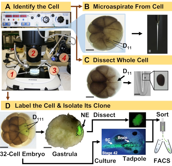
그림 1: 발달 중인 (개구리) 배아에서 세포 계통 유도 HRMS 단백질체학을 가능하게 하는 시공간적으로 확장 가능한 단백질체학. (A) 평행 전단 (4)에 의해 제어되는 제작된 마이크로피펫 (3)을 사용하여 확인된 세포(삽입물)의 주입을 위한 실체현미경(2)을 이용한 표본 (1)의 가시화. (B) 16-세포 배아에서 확인된 왼쪽D11 세포의 세포하 샘플링. (C) 16-세포 배아로부터 전체D11 세포의 해부. (D) 32세포 배아에서 왼쪽 및 오른쪽 D111 자손의 형광(녹색) 추적으로 위스트룰라에서 신경 외배엽(NE)의 해부(10단계) 및 FACS를 사용하여 올챙이에서 자손 조직 분리를 안내합니다. 스케일 바: 배아의 경우 200μm, 바이알의 경우 1.25mm. 수치는 참고 문헌 15,19,21,59의 허가를 받아 수정되었습니다. 이 그림의 더 큰 버전을 보려면 여기를 클릭하십시오.

여기에 제시된 프로토콜은 발달 중인 X. laevis 배아에서 확인된 세포/조직에서 많은 수의 단백질에 대한 HRMS 기반 정량화를 가능하게 합니다. 이 접근법은 정확한 세포 식별, 재현 가능한 세포 운명 지도 및 이 생물학적 모델 6,7,8에서 세포 계통을 추적하기 위한 확립된 방법론을 기반으로 합니다. 그림 1에서 볼 수 있듯이, 우리는 세포 내용물을 흡인하기 위해 전체 세포 해부 또는 모세관 마이크로 샘플링을 사용하여 단일 세포에서 프로테옴을 연구합니다. 세포의 혈통을 모니터링하면 세포가 위장 중에 조직을 형성함에 따라 프로테옴의 시공간 진화를 연구할 수 있습니다. 세포 자손은 형광 단백질(예: 녹색 형광 단백질 또는 GFP)에 대해 불활성 덱스트란 또는 mRNA에 접합된 형광단을 주입하여 형광을 표시합니다. 표지된 자손은 원하는 발달 시점에서 분리됩니다. gastrulation 동안, 단단히 클러스터링된 세포 클론은 해부에 의해 분리될 수 있습니다. 위축 후, 세포 클론은 이동 운동으로 인해 배아 내에 분포할 수 있으며 형광 활성화 세포 분류(FACS)에 의해 해리된 조직에서 분리될 수 있습니다. 이러한 세포 및 조직의 단백질은 분리를 위해 HPLC 또는 CE를 사용하고 식별을 위해 ESI 탠덤 HRMS를 사용하는 상향식 단백질체학을 통해 측정됩니다. 세포 계통 유도 HRMS 단백질체학은 배아 내에서 다양한 세포 크기와 계통으로 확장 가능하며 특이적이고 민감하며 정량적입니다. 여기에 표시된 선별된 예를 통해 이 프로토콜이 다양한 유형의 세포 및 세포 계통에 확장 가능하고 광범위하게 적응할 수 있음을 보여줍니다.

Figure 2
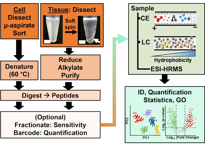
그림 2: 바이오분석 워크플로우. 미세 해부 및 모세관 흡인 또는 FACS는 세포 및 클론 단백질 함량의 샘플링을 용이하게 했습니다. 풍부한 난황 단백질의 고갈 및 모세관 전기영동(CE) 또는 나노 유동 액체 크로마토그래피(LC)에 의한 분리는 전기분무 이온화(ESI) 고분해능 질량분석법(HRMS)을 사용하여 식별(ID) 감도를 향상시킵니다. 정량화는 유전자 온톨로지(GO)에서 사용할 수 있는 정보와 함께 가설 기반 연구를 위한 새로운 정보를 제공하는 조절 장애를 드러냈습니다. 수치는 참고 문헌15의 허가를 받아 수정되었습니다. 이 그림의 더 큰 버전을 보려면 여기를 클릭하십시오.

Protocol

Xenopus laevis 성체 개구리의 인도적 유지 및 취급을 보장하는 모든 프로토콜은 메릴랜드 대학교 칼리지 파크의 기관 동물 관리 및 사용 위원회의 승인을 받았습니다(승인 번호 R-DEC-17-57 및 R-FEB-21-07). 1. 솔루션 준비 발생학의 경우1x, 0.5x 및 0.2x Steinberg’s solution(SS)을 준비한 다음 표준 프로토콜 12에 따라 멸균(120분 동안120°C)하여 ?…

Representative Results

이 프로토콜은 단일 세포에서 단백질과 X. laevis 배아에서 조직을 확립할 때 그 계통에 대한 연구를 가능하게 했습니다. 그림 1은 신경-조직 운명 세포와 배아에서 새로 유도된 신경 외배엽에서 단백질을 연구하기 위한 접근 방식의 이러한 적용 중 하나를 보여줍니다. 그림 1A에서 볼 수 있듯이 생물 분석 워크플로는 세포를 식별, 주입/흡인하고 표…

Discussion

이 프로토콜은 Xenopus 종의 배아에서 확인된 세포 계통에서 단백질 발현의 특성화를 가능하게 합니다. HRMS에서 비롯된 이 방법론은 분자 식별의 정교한 특이성, 분자 프로브 없이 다중 단백질 검출 기능(일반적으로 수백에서 수천 개의 서로 다른 단백질) 및 정량 기능을 결합합니다. 세포 및 발달 (신경) 생물학의 고전적인 도구 및 워크플로우에 대한 적응성은 척추동물 X. laevis 배아에?…

Disclosures

The authors have nothing to disclose.

Acknowledgements

배아 해리 및 FACS에 대한 귀중한 토론을 해주신 Jie Li (University of Maryland, College Park)에게 감사드립니다. 이 프로토콜에서 강조된 단백질체학 응용 분야를 예시하는 이전 연구에서 샘플 준비 및 데이터 수집에 도움을 준 Vi M. Quach와 Camille Lombard-Banek에게 감사드립니다. 이 연구의 일부는 수상 번호 IOS-1832968 CAREER(PN으로), 국립 보건원(National Institutes of Health)에서 수상 번호 R35GM124755(PN), 메릴랜드 대학교-국립 암 연구소 파트너십 프로그램(PN) 및 COSMOS Club Foundation 연구 상(ABB 및 LRP)의 지원을 받았습니다.

Materials

Acetonitrile (LC-MS-grade) Fisher Scientific A955
Agarose ThermoFisher Scientific R0492
Ammonium bicarbonate Fisher Scientific A643-500
Analytical Column Thermo Scientific 164941
Analytical microbalance Mettler-Toledo XSE105DU
Automatic peptide fractionation platform Agilent 1260 Infinity II
Borosilicate Capillaries Sutter Instruments Co. B100-50-10
Borosilicate Capillaries (for making Emmitters) Sutter Instruments B100-75-10
C18 spin columns (for desalting) ThermoFisher Scientific 89870
Camera ro monitor electrospray Edmund Optics Inc. EO-2018C
Combretastatin A4 Millipore Sigma C7744
Commercial CESI system AB SCIEX CESI
(Cyclohexylamino)-1-propanesulfonic acid (CAPS) VWR 97061-492
Cytochalasin D Millipore Sigma C8273
Dextran, Alexa Fluor 488; 10,000 MW, Anionic, Fixable ThermoFisher Scientific D22910
Diothiothreitol Fisher Scientific FERR0861
Dumont #5 Forceps Fine Science Tools 11252-30
EDTA Fisher Scientific AAJ62786AP
Epifluorescence light source Lumencore AURA III
Eppendorf LoBing microcentrifuge  tubes: protein Fisher Scientific 13-698-793
Formic acid (LC-MS-grade) Fisher Scientific A117-50
Freezer (-20 °C) Fisher Scientific 97-926-1 
Freezer (-80 °C) Thermo Scientific TSX40086A
Fused silica capillary Molex 1088150596
Heat Block Benchmark BSH300
High pressure liquid Chromatography System ThermoFisher Scientific Dionex Ultimate 3000 RSLC nanosystem
High voltage power supply Spellman CZE1000R
High-resolution Mass Spectrometer ThermoFisher Scientific Orbitrap Fusion Lumos Tribrid Mass Spectrometer
HPLC caps Thermo Scientific C4013-40A
HPLC Vials Thermo Scientific C4013-11
Illuminator e.g. Goosenecks Nikon C-FLED2
Ingenuity Pathway Analysis Qiagen
Iodoacetamide Fisher Scientific AC122275000
Methanol (LC-MS-grade) Fisher Scientific A456
Methanol (LC-MS-grade) Fisher Scientific A456-4
Microcapillary puller Suttor Instruments P-2000
Microinjector Warner Instrument, Handem, CT PLI-100A
Micropippette puller Sutter Instruments Co. P-1000
MS data analysis software, commercial ProteomeDiscoverer
MS data analysis software, opensource MaxQuant
non-idet 40 substitute Millipore Sigma 11754599001
Petri dish 60 mm and 80 mm Fisher Scientific S08184
Pierce 10 µL bed Zip-tips (for desalting) ThermoFisher Scientific 87782
Pierce bicinchoninic acid protein assay kit ThermoFisher Scientific 23225
Pierce quantitative colorimetric peptide assay ThermoFisher Scientific 23275
Pierce Trypsin Protease (MS Grade) Fisher Scientific PI90058
Protein LoBind vials Eppendorf 0030108434
, 0030108442
Refrigerated Centrifuge Eppendorf 5430R
Refrigerated Incubator Thermo Scientific PR505755R/3721
sodium isethionate Millipore Sigma 220078
sodium pyrophosphate Sigma Aldrich 221368-100G
Stainless steel BGE vial Custom-Built
Stainless steel sample vials Custom-Built
Stereomicroscope (objective 10x) Nikon SMZ 1270, SZX18
Sucrose VWR 97063-790
Syringe pumps (2) Harvard Apparatus 704506
Syringes (gas-tight): 500–1000 µL Hamilton 1750TTL
Transfer pipettes (Plastic, disposable) Fisher Scientific 13-711-7M
Trap Column Thermo Scientific 164750
Tris-HCl (1 M solution) Fisher Scientific AAJ22638AP
Vacuum concentrator capable of operation at 4–10 °C Labconco 7310022
Vortex-mixer Benchmark BS-VM-1000
Water (LC-MS-grade) Fisher Scientific W6
XYZ translation stage Thorlabs PT3
XYZ translation stage Custom-Built

References

  1. Shoemaker, L. D., Kornblum, H. I. Neural Stem Cells (NSCs) and Proteomics. Molecular & Cellular Proteomics. 15 (2), 344-354 (2016).
  2. Cervenka, J., et al. Proteomic characterization of human neural stem cells and their secretome during in vitro differentiation. Frontiers in Cellular Neuroscience. 14, 612560 (2021).
  3. Christian, J. L. Morphogen gradients in development: From form to function. Wiley Interdisciplinary Reviews. Developmental Biology. 1 (1), 3-15 (2012).
  4. Gurdon, J. B., Elsdale, T. R., M, F. Sexually mature individuals of Xenopus laevis from the transplantation of single somatic nuclei. Nature. 182, 64-65 (1958).
  5. Harland, R. M., Grainger, R. M. Xenopus research: metamorphosed by genetics and genomics. Trends in Genetics. 27 (12), 507-515 (2011).
  6. Moody, S. A. Fates of the blastomeres of the 16-cell stage Xenopus embryo. 发育生物学. 119 (2), 560-578 (1987).
  7. Moody, S. A. Fates of the blastomeres of the 32-cell stage Xenopus embryo. 发育生物学. 122 (2), 300-319 (1987).
  8. Dale, L., Slack, J. M. W. Fate map for the 32-cell stage of Xenopus laevis. Development. 99 (4), 527-551 (1987).
  9. Sun, L. L., et al. Single cell proteomics using frog (Xenopus laevis) blastomeres isolated from early stage embryos, which form a geometric progression in protein content. Analytical Chemistry. 88 (13), 6653-6657 (2016).
  10. Lombard-Banek, C., Moody, S. A., Nemes, P. Single-cell mass spectrometry for discovery proteomics: quantifying translational cell heterogeneity in the 16-cell frog (Xenopus) embryo. Angewandte Chemie-International Edition. 55 (7), 2454-2458 (2016).
  11. Zhang, Y. Y., Fonslow, B. R., Shan, B., Baek, M. C., Yates, J. R. Protein analysis by shotgun/bottom-up proteomics. Chemical Reviews. 113 (4), 2343-2394 (2013).
  12. Sive, H. L., Grainger, R. M., Harland, R. M. . Early development of Xenopus laevis: A laboratory manual. , (2000).
  13. Briggs, J. A., et al. The dynamics of gene expression in vertebrate embryogenesis at single-cell resolution. Science. 360 (6392), (2018).
  14. Gupta, M., Sonnett, M., Ryazanova, L., Presler, M., Wuhr, M., Vleminckx, K. Quantitative proteomics of xenopus embryos I, sample preparation. Xenopus. Methods in Molecular Biology. 1865, 175-194 (2018).
  15. Baxi, A. B., Lombard-Banek, C., Moody, S. A., Nemes, P. Proteomic characterization of the neural ectoderm fated cell clones in the Xenopus laevis embryo by high-resolution mass spectrometry. ACS Chemical Neuroscience. 9 (8), 2064-2073 (2018).
  16. Moody, S. A. Cell lineage analysis in Xenopus embryos. Methods in Molecular Biology. 135, 331-347 (2000).
  17. Sater, A. K., Moody, S. A. Using Xenopus to understand human diseases and developmental disorders. Genesis. 55 (1-2), 1-14 (2017).
  18. Lombard-Banek, C., Choi, S. B., Nemes, P., Allbritton, N. L., Kovarik, M. L. . Enzyme Activity in Single Cells. Methods in Enzymology. 628, 263-292 (2019).
  19. Lombard-Banek, C., Moody, S. A., Nemes, P. High-sensitivity mass spectrometry for probing gene translation in single embryonic cells in the early frog (Xenopus) embryo. Frontiers in Cell and Developmental Biology. 4, 11 (2016).
  20. Onjiko, R. M., Portero, E. P., Moody, S. A., Nemes, P. Microprobe capillary electrophoresis mass spectrometry for single-cell metabolomics in live frog (Xenopus laevis) embryos. Journal of Visualized Experiments: JoVE. (130), e56956 (2017).
  21. Lombard-Banek, C., Moody, S. A., Manzin, M. C., Nemes, P. Microsampling capillary electrophoresis mass spectrometry enables single-cell proteomics in complex tissues: developing cell clones in live Xenopus laevis and zebrafish embryos. Analytical Chemistry. 91 (7), 4797-4805 (2019).
  22. Klein, S. L. The first cleavage furrow demarcates the dorsal-ventral axis in Xenopus embryos. 发育生物学. 120 (1), 299-304 (1987).
  23. Karimi, K., et al. Xenbase: a genomic, epigenomic and transcriptomic model organism database. Nucleic Acids Research. 46 (1), 861-868 (2018).
  24. Kakebeen, A. D., Chitsazan, A. D., Wills, A. E. Tissue disaggregation and isolation of specific cell types from transgenic Xenopus appendages for transcriptional analysis by FACS. Developmental Dynamics. 250 (9), 1381-1392 (2021).
  25. Garcia, B. A. What does the future hold for top down mass spectrometry. Journal of the American Society for Mass Spectrometry. 21 (2), 193-202 (2010).
  26. Toby, T. K., Fornelli, L., Kelleher, N. L. Progress in top-down proteomics and the analysis of proteoforms. Annual Review of Analytical Chemistry. (Palo Alto Calif). 9 (1), 499-519 (2016).
  27. Zhang, Z. B., Dubiak, K. M., Huber, P. W., Dovichi, N. J. Miniaturized filter-aided sample preparation (MICRO-FASP) method for high throughput, ultrasensitive proteomics sample preparation reveals proteome asymmetry in Xenopus laevis Embryos. Analytical Chemistry. 92 (7), 5554-5560 (2020).
  28. Wisniewski, J. R., Becher, D. . Microbial Proteomics: Methods and Protocols.Methods in Molecular Biology. 1841, 3-10 (2018).
  29. Hughes, C. S., et al. Single-pot, solid-phase-enhanced sample preparation for proteomics experiments. Nature Protocols. 14 (1), 68-85 (2019).
  30. Zhu, Y., et al. Nanodroplet processing platform for deep and quantitative proteome profiling of 10-100 mammalian cells. Nature Communications. 9, 882 (2018).
  31. Wessel, D., Flugge, U. I. A method for the quantitative recovery of protein in dilute-solution in the presence of detergents and lipids. Analytical Biochemistry. 138 (1), 141-143 (1984).
  32. Jiang, L., He, L., Fountoulakis, M. Comparison of protein precipitation methods for sample preparation prior to proteomic analysis. Journal of Chromatography A. 1023 (2), 317-320 (2004).
  33. Hildonen, S., Halvorsen, T. G., Reubsaet, L. Why less is more when generating tryptic peptides in bottom-up proteomics. Proteomics. 14 (17-18), 2031-2041 (2014).
  34. Budnik, B., Levy, E., Harmange, G., Slavov, N. SCoPE-MS: mass spectrometry of single mammalian cells quantifies proteome heterogeneity during cell differentiation. Genome Biology. 19, 161 (2018).
  35. Drouin, N., et al. Capillary electrophoresis-mass spectrometry at trial by metabo-ring: effective electrophoretic mobility for reproducible and robust compound annotation. Analytical Chemistry. 92 (20), 14103-14112 (2020).
  36. Sun, L. L., Zhu, G. J., Zhang, Z. B., Mou, S., Dovichi, N. J. Third-generation electrokinetically pumped sheath-flow nanospray interface with improved stability and sensitivity for automated capillary zone electrophoresis-mass spectrometry analysis of complex proteome digests. Journal of Proteome Research. 14 (5), 2312-2321 (2015).
  37. DeLaney, K., Sauer, C. S., Vu, N. Q., Li, L. J. Recent advances and new perspectives in capillary electrophoresis-mass spectrometry for single cell "omics". Molecules. 24 (1), 21 (2019).
  38. Nemes, P., Rubakhin, S. S., Aerts, J. T., Sweedler, J. V. Qualitative and quantitative metabolomic investigation of single neurons by capillary electrophoresis electrospray ionization mass spectrometry. Nature Protocols. 8 (4), 783-799 (2013).
  39. Choi, S. B., Zamarbide, M., Manzini, M. C., Nemes, P. Tapered-tip capillary electrophoresis nano-electrospray ionization mass spectrometry for ultrasensitive proteomics: the mouse cortex. Journal of the American Society for Mass Spectrometry. 28 (4), 597-607 (2017).
  40. Pino, L. K., Rose, J., O’Broin, A., Shah, S., Schilling, B. Emerging mass spectrometry-based proteomics methodologies for novel biomedical applications. Biochemical Society Transactions. 48 (5), 1953-1966 (2020).
  41. Chen, C., Hou, J., Tanner, J. J., Cheng, J. L. Bioinformatics methods for mass spectrometry-based proteomics data analysis. International Journal of Molecular Sciences. 21 (8), 25 (2020).
  42. Peshkin, L., et al. On the relationship of protein and mRNA dynamics in vertebrate embryonic development. Developmental Cell. 35 (3), 383-394 (2015).
  43. Cox, J., et al. Accurate proteome-wide label-free quantification by delayed normalization and maximal peptide ratio extraction, termed MaxLFQ. Molecular & Cellular Proteomics. 13 (9), 2513-2526 (2014).
  44. Gygi, S. P., et al. Quantitative analysis of complex protein mixtures using isotope-coded affinity tags. Nature Biotechnology. 17 (10), 994-999 (1999).
  45. Thompson, A., et al. Tandem mass tags: A novel quantification strategy for comparative analysis of complex protein mixtures by MS/MS. Analytical Chemistry. 75 (8), 1895-1904 (2003).
  46. Mi, H. Y., et al. PANTHER version 16: a revised family classification, tree-based classification tool, enhancer regions and extensive api. Nucleic Acids Research. 49, 394-403 (2021).
  47. Schmidt, E., et al. . On the Move Federated Workshops. , 710-719 (2006).
  48. Deutsch, E. W., et al. Trans-Proteomic pipeline, a standardized data processing pipeline for large-scale reproducible proteomics informatics. Proteomics Clinical Applications. 9 (7-8), 745-754 (2015).
  49. Tyanova, S., et al. The Perseus computational platform for comprehensive analysis of (prote)omics data. Nature Methods. 13 (9), 731-740 (2016).
  50. Demsar, J., et al. Orange: Data mining toolbox in Python. Journal of Machine Learning Research. 14, 2349-2353 (2013).
  51. Oberg, A. L., Vitek, O. Statistical design of quantitative mass spectrometry-based proteomic experiments. Journal of Proteome Research. 8 (5), 2144-2156 (2009).
  52. Jensen, L. J., et al. STRING 8 – a global view on proteins and their functional interactions in 630 organisms. Nucleic Acids Research. 37, 412-416 (2009).
  53. Schweppe, D. K., Huttlin, E. L., Harper, J. W., Gygi, S. P. BioPlex display: an interactive suite for large-scale AP-MS protein-protein interaction data. Journal of Proteome Research. 17 (1), 722-726 (2018).
  54. Hornbeck, P. V., et al. PhosphoSitePlus, 2014: mutations, PTMs and recalibrations. Nucleic Acids Research. 43, 512-520 (2015).
  55. Letunic, I., Khedkar, S., Bork, P. SMART: recent updates, new developments and status in 2020. Nucleic Acids Research. 49, 458-460 (2021).
  56. Lombard-Banek, C., et al. In vivo subcellular mass spectrometry enables proteo-metabolomic single-cell systems biology in a chordate embryo developing to a normally behaving tadpole (X. laevis). Angewandte Chemie-International Edition. 60 (23), 12852-12858 (2021).
  57. Lombard-Banek, C., Reddy, S., Moody, S. A., Nemes, P. Label-free quantification of proteins in single embryonic cells with neural fate in the cleavage-stage frog (Xenopus laevis) embryo using capillary electrophoresis electrospray ionization high-resolution mass spectrometry (CE-ESI-HRMS). Molecular & Cellular Proteomics. 15 (8), 2756-2768 (2016).
  58. Saha-Shah, A., et al. Single cell proteomics by data-independent acquisition to study embryonic asymmetry in Xenopus laevis. Analytical Chemistry. 91 (14), 8891-8899 (2019).
  59. Onjiko, R. M., Portero, E. P., Moody, S. A., Nemes, P. In situ microprobe single-cell capillary electrophoresis mass spectrometry: metabolic reorganization in single differentiating cells in the live vertebrate (Xenopus laevis) embryo. Analytical Chemistry. 89 (13), 7069-7076 (2017).
  60. Perez-Riverol, Y., et al. The PRIDE database and related tools and resources in 2019: improving support for quantification data. Nucleic Acids Research. 47, 442-450 (2019).
check_url/cn/63586?article_type=t

Play Video

Cite This Article
Baxi, A. B., Pade, L. R., Nemes, P. Cell-Lineage Guided Mass Spectrometry Proteomics in the Developing (Frog) Embryo. J. Vis. Exp. (182), e63586, doi:10.3791/63586 (2022).

View Video