Summary

चूहे में एक साधारण क्रिटिकल आकार ऊरु दोष मॉडल

Published: March 15, 2015
doi:

Summary

Animal models are frequently employed to mimic serious bone injury in biomedical research. Due to their small size, establishment of stabilized bone lesions in mice are beyond the capabilities of most research groups. Herein, we describe a simple method for establishing and analyzing experimental femoral defects in mice.

Abstract

While bone has a remarkable capacity for regeneration, serious bone trauma often results in damage that does not properly heal. In fact, one tenth of all limb bone fractures fail to heal completely due to the extent of the trauma, disease, or age of the patient. Our ability to improve bone regenerative strategies is critically dependent on the ability to mimic serious bone trauma in test animals, but the generation and stabilization of large bone lesions is technically challenging. In most cases, serious long bone trauma is mimicked experimentally by establishing a defect that will not naturally heal. This is achieved by complete removal of a bone segment that is larger than 1.5 times the diameter of the bone cross-section. The bone is then stabilized with a metal implant to maintain proper orientation of the fracture edges and allow for mobility.

Due to their small size and the fragility of their long bones, establishment of such lesions in mice are beyond the capabilities of most research groups. As such, long bone defect models are confined to rats and larger animals. Nevertheless, mice afford significant research advantages in that they can be genetically modified and bred as immune-compromised strains that do not reject human cells and tissue.

Herein, we demonstrate a technique that facilitates the generation of a segmental defect in mouse femora using standard laboratory and veterinary equipment. With practice, fabrication of the fixation device and surgical implantation is feasible for the majority of trained veterinarians and animal research personnel. Using example data, we also provide methodologies for the quantitative analysis of bone healing for the model.

Introduction

यह इलाज भंग के साथ उन रोगियों के लिए शल्य चिकित्सा, 500,000 प्रक्रियाओं बोन ग्राफ्ट 2 का उपयोग शामिल है। अमेरिका की आबादी का आधा 65 1 साल की उम्र से एक फ्रैक्चर का अनुभव है कि अनुमान है और इस संख्या में एक तेजी से उम्र बढ़ने की आबादी 3 के साथ वृद्धि की उम्मीद है । हड्डी पूरी तरह से scarring के बिना चंगा करने के लिए क्षमता है कि कुछ अंगों में से एक है हालांकि प्रक्रिया 3,4 विफल रहता है, जहां उदाहरण हैं। हालात और इलाज की गुणवत्ता पर निर्भर करता है, लंबे समय से अस्थि भंग का 2-30% गैर संघ 3,5, जिसके परिणामस्वरूप में असफल। परिभाषा, pseudoarthrosis, आकार-महत्वपूर्ण या गैर संघ की हड्डी की चोटों पर कुछ बहस बनी हुई है जबकि आम तौर पर इस विषय 6 के प्राकृतिक जीवनकाल में ठीक नहीं है कि एक चोट को दर्शाता है। प्रयोगात्मक प्रयोजनों के लिए, इस अवधि के एक औसत आकार की हड्डी की चोट का पूर्ण उपचार के लिए आवश्यक औसत समय के लिए छोटा है। गैर संघ हड्डी घावों NUM के लिए होते हैंerous कारणों से है, लेकिन प्रमुख कारकों की वजह से बीमारी या 7 साल की उम्र के लिए एक गंभीर रूप से आकार की खाई, संक्रमण, गरीब एंजियोजिनेसिस, तंबाकू के इस्तेमाल, या हिचकते osteoregenerative क्षमता में जिसके परिणामस्वरूप चरम आघात शामिल हैं। गैर यूनियनों सफलतापूर्वक इलाज कर रहे हैं, भले ही यह चोट के प्रकार और 8 कार्यरत दृष्टिकोण पर निर्भर करता है, प्रक्रिया के अनुसार 60,000 डॉलर की अतिरिक्त में खर्च कर सकते हैं।

उदारवादी मामलों में, ऑटोलॉगस बोन ग्राफ्टिंग कार्यरत है। इस रणनीति के चोट के स्थल पर एक दाता साइट और आरोपण से हड्डी की वसूली शामिल है। इस दृष्टिकोण अत्यंत प्रभावी है, उपलब्ध दाता व्युत्पन्न हड्डी की मात्रा सीमित है और प्रक्रिया कई रोगियों 9,10 में लगातार दर्द में जो परिणाम एक अतिरिक्त सर्जरी, शामिल है। इसके अलावा, ऑटोलॉगस बोन ग्राफ्ट की प्रभावकारिता रोगी के स्वास्थ्य निर्भर है। सिंथेटिक सामग्री या संसाधित शव हड्डी से बना अस्थि के विकल्प 11-13 बहुतायत से उपलब्ध हैं, लेकिन वे हागरीब मेजबान कोशिका आसंजन गुण, कम osteoconductivity, और प्रतिरक्षा अस्वीकृति 14 के लिए संभावित सहित महत्वपूर्ण सीमाओं, ve। सुरक्षित, प्रभावी और व्यापक रूप से उपलब्ध हैं कि हड्डी पुनर्जनन प्रौद्योगिकियों के लिए एक तत्काल आवश्यकता इसलिए नहीं है।

हड्डी पुनर्योजी रणनीतियों में सुधार करने की क्षमता का परीक्षण पशुओं में गंभीर हड्डी आघात नकल करने की क्षमता पर निर्भर है, लेकिन बड़े हड्डी घावों की पीढ़ी और स्थिरीकरण तकनीकी रूप से चुनौती दे रहा है। ज्यादातर मामलों में, गंभीर लंबी हड्डी आघात स्वाभाविक रूप से ठीक नहीं होगा कि एक दोष की स्थापना के द्वारा प्रयोगात्मक मजाक उड़ाया जाता है। यह प्रजाति 15 के साथ अलग-अलग हो सकता है, यह हड्डी पार अनुभाग 16 के बड़े से 1.5 गुना व्यास है कि एक हड्डी खंड के पूरी तरह हटाने के द्वारा हासिल की है। हड्डी तो फ्रैक्चर किनारों की उचित उन्मुखीकरण को बनाए रखने और गतिशीलता के लिए अनुमति देने के लिए एक धातु प्रत्यारोपण के साथ स्थिर है। कारण उनके छोटे आकार और की कमजोरी कोउनकी लंबी हड्डियों, चूहों में इस तरह के घावों की स्थापना के सबसे अनुसंधान समूहों की क्षमताओं से परे हैं। जैसे, लंबी हड्डी दोष मॉडल चूहों और बड़े जानवरों तक ही सीमित हैं। फिर भी, चूहों वे आनुवंशिक रूप से संशोधित और मानव कोशिकाओं और ऊतकों को अस्वीकार नहीं करते कि प्रतिरक्षा समझौता किया उपभेदों के रूप में पैदा किया जा सकता है कि में महत्वपूर्ण अनुसंधान फायदे बनती हैं।

मानव कोशिका आधारित अनुप्रयोगों के लिए, प्रतिरक्षा-समझौता किया चूहों वे physiologically घर के लिए आसान है, अच्छी तरह से विशेषता हैं क्योंकि के साथ काम करने के लिए आकर्षक हैं, लागत प्रभावी, और आसानी से histologically radiologically और विश्लेषण किया। सर्वोच्च महत्व का प्रतिरक्षा समझौता किया चूहों मानव सहित विभिन्न प्रजातियों से कोशिकाओं को अस्वीकार नहीं है। अपने छोटे आकार भी कोशिकाओं या आर्थोपेडिक अनुप्रयोगों में प्रयोगात्मक scaffolds के संस्करणों की बहुत छोटी संख्या के परीक्षण के सभी देता है। कई murine आर्थोपेडिक मॉडल हड्डी स्थिरता 17,18 के विभिन्न डिग्री बर्दाश्त कि सूचना दी गई है। उन systeendochondral चिकित्सा 19 सूचित किया गया है, हालांकि इस तरह के बाहरी fixators और ताला लगा प्लेटों के रूप में स्थिरता का बहुत ही उच्च स्तर, में परिणाम है कि एमएस मुख्य रूप से intramembranous हड्डी बन जाना द्वारा चंगा। इसके विपरीत, इस तरह के unfixed या आंशिक रूप से निश्चित दिमाग़ी पिन को रोजगार के रूप में उन कुछ सूक्ष्म और / या मैक्रो-गति, की अनुमति उन है कि आम तौर पर endochondral हड्डी बन जाना 20,21 की एक विशेषता के साथ चंगा। विलंबित संघ या लंबी हड्डी की गैर संघ दोष की वजह से आवश्यक स्थिरीकरण के अतिरिक्त स्तर तक चूहों में प्राप्त करने के लिए विशेष रूप से कठिन हैं। हालांकि, दृष्टिकोण के एक नंबर इंटरलॉकिंग नाखून के साथ मस्तिष्क का पिन, ताला लगा प्लेट्स और बाहरी fixators 22 सहित सूचित किया गया है। इन प्रणालियों को आम तौर पर अच्छी तरह से काम करते हैं, लेकिन उनकी जटिल डिजाइन वे स्थापित करने के लिए तकनीकी रूप से चुनौतीपूर्ण हो सकता दी। उदाहरण के लिए, गार्सिया एट अल। 23 चूहों में उपयोग के लिए एक सुंदर इंटरलॉकिंग पिन प्रणाली तैयार है, लेकिन प्रक्रिया दो अलग-अलग स्थल पर चीरों शामिलs और फीमर की व्यापक संशोधन पिन समायोजित करने के लिए। इन प्रक्रियाओं के एक विदारक माइक्रोस्कोप के तहत प्रदर्शन किया गया।

इस के साथ साथ, हम एक 3 मिमी हड्डी घाटे का समापन रोकने के लिए और भी दोष के मूल किनारों को चित्रित करने के लिए डिजाइन एक केंद्रीय कॉलर के साथ एक सरल और्विक दिमाग़ी पिन का वर्णन है। पिन हड्डी खुद के लिए तय नहीं किया गया है, जबकि पर्याप्त हस्तक्षेप में दिमाग़ी गुहा परिणामों की पिन व्यास और Reaming की सटीक नौकरशाही का आकार घटाने मरोड़ गति (चित्रा 1) को कम से कम करने के लिए। जन्मजात उम्र, लिंग और तनाव-मिलान चूहों का चयन सावधानी के साथ, परिणाम आसानी से radiologically मूल्यांकन किया जा सकता है, जो एक बेहद प्रतिलिपि प्रस्तुत करने योग्य hypertrophic गैर uniondefect 22 है। इसके अलावा ब्याज के क्षेत्रों reproducibly डी नोवो हड्डी गठन और histomorphological मापदंडों की माप के लिए सूक्ष्म गणना टोमोग्राफी (μCT) के बाद से परिभाषित किया जा सकता है। पिन आसानी से उपलब्ध उपकरणों का उपयोग हमारी प्रयोगशाला में prototyped थे।

चित्र 1
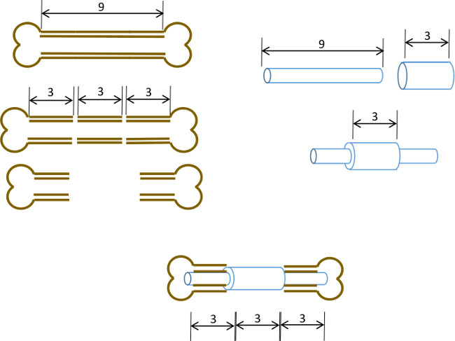
चित्रा 1: प्रायोगिक सिद्धांत कमानी दोष मॉडल के ढांचे के सारांश।। एक 9-10 मिमी murine फीमर के मध्य 3 मिमी खंड (बाएं) शल्य चिकित्सा excised है। एक 3 मिमी लंबा, 19 गेज शल्य स्टील ट्यूब सटीक केंद्र (दाएं) में लंबे समय से एक 9 एमएम, 22 ग्राम स्टेनलेस स्टील ट्यूब पर पारित किया है और चिपकने के साथ तय हो गई है। परिणामी पिन हड्डी के 3 मिमी खंड की जगह 19 जी कॉलर (नीचे, केंद्र) के साथ फीमर के शेष प्रॉक्सिमल और बाहर भाग के दिमाग़ी नहरों में लगाया जाता है।

Protocol

नोट: इस प्रोटोकॉल जैक्सन प्रयोगशालाओं से हासिल कर ली महिला नग्न (एन यू / जम्मू) चूहों (18-25 ग्राम, 6 सप्ताह) के लिए बनाया गया है। चूहों के तनाव शरीर रचना और विकास दर के मामले में थोड़ा भिन्न बाद से, हम पिन के निर…

Representative Results

चूहे आम तौर पर चेतना की वसूली और हिंद अंग गतिशीलता संज्ञाहरण की वापसी के बाद 5-10 मिनट। पहले 5 दिनों के दौरान, यह व्यक्तिगत रूप से चूहों को घर और अंग से अधिक उपयोग को रोकने के लिए पर्यावरण संवर्धन शुरू करने ?…

Discussion

इस के साथ साथ, हम मानक प्रयोगशाला और पशु चिकित्सा उपकरण का उपयोग कर murine फीमर की एक महत्वपूर्ण आकार पिन स्थिर दोष उत्पन्न करने के लिए एक सरल विधि का वर्णन। पिन और शल्य प्रक्रिया ही के विधानसभा अभ्यास की आव…

Disclosures

The authors have nothing to disclose.

Acknowledgements

हम इस तकनीक के विकास के दौरान उनके अमूल्य सलाह और सहायता के लिए, तुलनात्मक चिकित्सा, मंदिर, टेक्सास के स्कॉट एंड व्हाइट अस्पताल विभाग में कर्मचारियों और पशु चिकित्सकों को धन्यवाद। इस काम पुनर्योजी चिकित्सा कार्यक्रम फंड के लिए संस्थान, स्कॉट एंड व्हाइट RGP अनुदान # 90,172, एनआईएच 2P40RR017447-07 और एनआईएच R01AR066033-01 (NIAMS) द्वारा वित्त पोषित में भाग गया था। हम पांडुलिपि अशुद्धि के लिए डॉ थीं Zeitouni धन्यवाद।

Materials

Name of Equipment/Material* Company Catalog or model Notes
Pin Assembly
Dremel rotary tool Dremel 8220 or equivalent
Heavy duty cut off wheel Dremel 420
Surgical tubing 19G Small Parts (Amazon) B000FMZ8LY OD 1.07mm, ID 0.889mm
Surgical tubing 21G Small Parts (Amazon) B000FMZ8YQ OD 0.82mm, ID 0.635mm
Surgical tubing 22G Small Parts (Amazon) B000FMYLZS OD 0.719mm, ID 0.502mm
Surgical tubing 23G Small Parts (Amazon) B000FN0SY0 OD 0.643mm, ID 0.444mm
Cyanoacrylate adhesive Loctite 1365882
Emery disc Dremel 413
Rubber polishing point Dremel 462
Felt polishing disc Dremel 414
Gelatin sponge Surgifoam/Ethicon 1974
Punch biopsy cutter Miltex 33-34
Surgery/post-operative
Warm pad and circulator pump Stryker/Thermocare TP700, TP700C, TPP722
Coverage quaternary spray Steris 1429-77
Bead sterilizer Germinator/CellPoint Scentific Germinator 500
Anesthesia system VetEquip Inc 901806 or 901807/901809
Isofluorane anesthetic VETone/MWI 501017, 502017
Surgical disinfectant Chloraprep/CareFusion 260449
Surgical tools Fine Science Tools various recommend German made
Face protection Splash Shield 4505
Rechargable high speed drill Fine Science Tools 18000-17
Diamond cutting wheel Strauss Diaiond 361.514.080HP
Absorbable sutures  Covidien UM-213
Outer sutures Ethicon 668G or equivalent
Vetbond 3M 1469SB or equivalent
Hydration gel Clear H2O 70-01-1082
Diet gel Clear H2O 72-01-1062
Buprenorphine Reckitt and Benckser 12496-0757-01 controlled substance
Mouse igloos Bio Serv K3328, 3570,3327
Euthanasia cocktail Euthasol/Virbac 710101 controlled substance
Analysis
Live animal imager  Orthoscan FD Pulse or equivalent
Micro-CT unit and software Bruker Skyscan1174 or equivalent
Sealing film/Parafilm M VWR or Fisher 100501-338, S37441
*Generic sources are suitable for all other items such as gause, drapes, protective clothing, animal care equipment.

References

  1. Brinker, M. R., O’Connor, D. P. The incidence of fractures and dislocations referred for orthopaedic services in a capitated population. J Bone Joint Surg Am. 86, 290-297 (2004).
  2. Cheung, C. The future of bone healing. Clin Podiatr Med Surg. 22, 631-641 (2005).
  3. Rosemont, I. L. . United States Bone and Joint Decade: The burden of musculoskeletal diseases and musculoskeletal injuries. , (2008).
  4. Tzioupis, C., Giannoudis, P. V. Prevalence of long-bone non-unions. Injury. 38, S3-S9 (2007).
  5. Marsh, D. Concepts of fracture union, delayed union, and nonunion. Clin Orthop Relat Res. , S22-S30 (1998).
  6. Spicer, P. P., et al. Evaluation of bone regeneration using the rat critical size calvarial defect. Nat Protoc. 7, 1918-1929 (2012).
  7. Green, E., Lubahn, J. D., Evans, J. Risk factors, treatment, and outcomes associated with nonunion of the midshaft humerus fracture. J Surg Orthop Adv. 14, 64-72 (2005).
  8. Kanakaris, N. K., Giannoudis, P. V. The health economics of the treatment of long-bone non-unions. Injury. 38, S77-S84 (2007).
  9. Dimitriou, R., Mataliotakis, G. I., Angoules, A. G., Kanakaris, N. K., Giannoudis, P. V. Complications following autologous bone graft harvesting from the iliac crest and using the RIA: a systematic review. Injury. 42, S3-S15 (2011).
  10. Boer, H. H. The history of bone grafts. Clin Orthop Relat Res. , 292-298 (1988).
  11. Aro, H. T., Aho, A. J. Clinical use of bone allografts. Ann Med. 25, 403-412 (1993).
  12. Burstein, F. D. Bone substitutes. Cleft Palate Craniofac. J. 37, 1-4 (2000).
  13. Kao, S. T., Scott, D. D. A review of bone substitutes. Oral Maxillofac Surg Clin North Am. 19, 513-521 (2007).
  14. Boden, S. D. Overview of the biology of lumbar spine fusion and principles for selecting a bone graft substitute. Spine. (Phila Pa 1976). 27, S26-S31 (1976).
  15. Hollinger, J. O., Kleinschmidt, J. C. The critical size defect as an experimental model to test bone repair materials). J Craniofac Surg. 1, 60-68 (1990).
  16. Key, J. The effect of local calcium depot on osteogenesis and healing of fractures. J. Bone Joint Surg. (Am). 16, 176-184 (1934).
  17. Holstein, J. H., et al. Advances in the establishment of defined mouse models for the study of fracture healing and bone regeneration). J Orthop Trauma. 23, S31-S38 (2009).
  18. Histing, T., et al. Small animal bone healing models: standards, tips, and pitfalls results of a consensus meeting. Bone. 49, 591-599 (2011).
  19. Cheung, K. M., et al. An externally fixed femoral fracture model for mice. J Orthop Res. 21, 685-690 (2003).
  20. Hiltunen, A., Vuorio, E., Aro, H. T. A standardized experimental fracture in the mouse tibia. J Orthop Res. 11, 305-312 (1993).
  21. Manigrasso, M. B., O’Connor, J. P. Characterization of a closed femur fracture model in mice. J Orthop Trauma. 18, 687-695 (2004).
  22. Garcia, P., et al. Rodent animal models of delayed bone healing and non-union formation: a comprehensive review. Eur Cell Mater. 26, 1-12 (2013).
  23. Garcia, P., et al. Development of a reliable non-union model in mice. J Surg Res. 147, 84-91 (2008).
  24. Flecknell, P. A. The relief of pain in laboratory animals. Lab Anim. 18, 147-160 (1984).
  25. . . Guidelines on the Euthanasia of Animals. , (2013).
  26. Neill, K. R., et al. Micro-computed tomography assessment of the progression of fracture healing in mice. Bone. 50, 1357-1367 (2012).
  27. Bagi, C. M., et al. The use of micro-CT to evaluate cortical bone geometry and strength in nude rats: correlation with mechanical testing, pQCT and DXA. Bone. 38, 136-144 (2006).
  28. Hadjiargyrou, M., et al. Transcriptional profiling of bone regeneration. Insight into the molecular complexity of wound repair. J Biol Chem. 277, 30177-30182 (2002).
  29. Clough, B. H., et al. Bone regeneration with osteogenically enhanced mesenchymal stem cells and their extracellular matrix proteins. J Bone Miner Res. , (2014).
  30. Lu, C., et al. Cellular basis for age-related changes in fracture repair. J Orthop Res. 23, 1300-1307 (2005).
  31. Jepsen, K. J., et al. Genetic variation in the patterns of skeletal progenitor cell differentiation and progression during endochondral bone formation affects the rate of fracture healing. J Bone Miner Res. 23, 1204-1216 (2008).
  32. Thayer, T. C., Wilson, S. B., Mathews, C. E. Use of nonobese diabetic mice to understand human type 1 diabetes. Endocrinol Metab Clin North Am. 39, 541-561 (2010).
  33. Jee, W. S., Yao, W. Overview: animal models of osteopenia and osteoporosis. J Musculoskelet Neuronal Interact. 1, 193-207 (2001).
check_url/52368?article_type=t

Play Video

Cite This Article
Clough, B. H., McCarley, M. R., Gregory, C. A. A Simple Critical-sized Femoral Defect Model in Mice. J. Vis. Exp. (97), e52368, doi:10.3791/52368 (2015).

View Video