Summary

解剖学启发的三维微组织工程神经网络用于神经系统重建,调制和建模

Published: May 31, 2017
doi:

Summary

该手稿详细描述了微组织工程神经网络的制造:三维微米大小的构建体,包括跨越聚集的神经元群体的长比例的轴突区域,其包裹在管状水凝胶中。这些生活支架可以作为功能性继电器来重建或调节神经回路,或者作为模拟灰白色神经解剖学的生物学检验床。

Abstract

由于抑制环境和神经发生能力有限,功能恢复很少发生在中枢神经系统(CNS)中的损伤或疾病诱导的变性之后。我们正在开发一种同时解决损伤的CNS内的神经元和轴突途径损失的策略。该手稿提出了微组织工程神经网络(微型TENN)的制造方案,由神经元组成的可植入构造物和跨越细胞外基质(ECM)腔的对准轴突束,其预先形成的直径为数百微米的水凝胶圆柱体可以延伸厘米长度。神经元聚集体被划分为三维包围的极端,并被轴突突起所跨越。微型TENN作为CNS重建的策略是独一无二的,模拟了脑连接体细胞结构的方面,并为潜在的网络替代提供了手段。神奈轮状聚集体可能与宿主组织突触形成新的功能性继电器,以恢复和/或调节丢失或损坏的电路。这些构建体也可以作为能够利用细胞迁移和轴索寻路的发育机制的再生“活支架”,基于再生状态提供协同的结构和可溶性线索。将微型TENN通过将液体水凝胶倒入含有纵向中心针的圆柱形模具中来制造。一旦水凝胶凝胶化,就去除针头,留下中空微柱。将ECM溶液加入腔内以提供适合于神经元粘附和轴突生长的环境。机械地聚集离解的神经元用于在微柱的一端或两端精确播种。该方法可靠地产生具有长突出的轴突束的自足的微型构造,其可以重现脑神经解剖学的特征。突触动物非标记和遗传编码的钙指标表明微TENN具有广泛的突触分布和内在的电活动。因此,微型TENN代表脑途径靶向神经外科重建的有希望的策略,也可作为生物学模型应用于体外研究神经生物学现象

Introduction

中枢神经系统疾病和疾病(CNS)的常见特征,如创伤性脑损伤(TBI),脊髓损伤(SCI),中风,阿尔茨海默病和帕金森病,是轴突途径和神经元细胞的断开损失1,2,3,4,5,6 。例如,当缺血性中风未经治疗时,估计轴突以每分钟轴突7英里的速度丢失5 。在TBI的情况下,每年约有170万人在美国经历,轴突变性可能在创伤后几年可能继续发生,因为初始损伤导致长期的神经变性状态4 。加剧这些有害影响,中枢神经系统受到严重限制城市再生1,7,8,9。受伤后,出现抑制环境,其特征在于缺乏远距离靶点的指导性指导,阻止神经突生长的髓磷脂相关抑制剂的存在,以及由反应性星形胶质细胞形成胶质瘢痕8,10,11,12。胶质瘢痕可作为再生的生化和物理障碍,硫酸软骨素蛋白多糖等分子阻碍轴突长出8,11。此外,即使在成人CNS中发现了神经干细胞,新神经元的产生也是有限的,因为在嗅球中仅发现了神经发生的一致证据,海马脊髓区域,脑室周围区域和脊髓中央管道13,14 。这些障碍阻碍了损伤或疾病后遗失的神经元和白质结构的功能恢复,导致这些病症经常改变生命和延长的效果。

尽管成人中枢神经系统缺乏再生能力,但是已经证明,如果向宿主神经元15,16,17,18提供足够的环境线索,则轴突再生是可能的。研究人员尝试提供和操纵生长因子( 神经生长因子,表皮生长因子,神经胶质依赖性生长因子和神经营养因子-3)和其他指导分子,以刺激可塑性和轴突再生14 / sup> 18,19 。即使这些研究已经证实成年轴突能够响应生长因子,这些策略受到血脑屏障的低渗透性和促进再生所需的特定空间和时间梯度的限制14,18,19。其他方法依赖于CNS神经元中再生相关转录因子的过度活化。例如,Stat3转录因子的过度表达刺激视神经中的轴突再生20 。然而,生物分子递送和转录因子的过表达都不能代替丢失的神经元群体。基于细胞的策略主要集中在将神经干细胞(NSCs)移植到CNS中,利用其替代CNS神经元的能力,释放营养因子,并支持在损伤后发生的神经发生的尝试17 。尽管如此,仍然存在阻碍这种方法的紧迫挑战,包括移植的神经细胞存活,与主机整合的阻碍能力,并且在空间上保持受限制的区域6,14,17,21。此外,单独的细胞传递不能恢复受损或丢失的轴突途径的细胞结构。解决细胞和药物/化学品输送策略面临的问题的另一种方法是将这些方法与生物材料14,22,23的使用相结合。生物材料如水凝胶能够模拟细胞外基质(ECM)的生物化学和物理性质,有助于细胞递送d保留在受伤区域内,并传递生长因子和其他生物活性分子与受控释放22 。这些基于生物材料的策略的有吸引力的特征已经导致在支架移植到损伤区域24,25,26,27,28,29,30之后体内轴突再生的证据。然而,无细胞生物材料策略不能代替丢失的神经元群体;当用作神经元,胶质细胞或神经元前体细胞的递送载体时,生物材料不能重建长距离轴突网络。开发解决轴突途径退化和与CNS损伤和疾病相关的神经元损失的方法的挑战仍然是<sup class =“xref”> 31。

我们的研究小组以前报告了可植入的微组织工程神经网络(micro-TENNs)的发展,这是一种由活性支架组成的神经细胞体,其限于琼脂糖水凝胶-EGC微柱的一端或两端,其中对齐的轴突束延伸穿过该三维(3D)包装1,10,31,32的整个内部。这种技术和以前的方法之间的主要区别之一是微TENN 的细胞结构在体外完全产生并在之后33,34,35,36,37,38 sup class =“xref”> 39,40,41。 体外制备提供了广泛的空间和时间控制细胞表型和取向,机械/物理性质,生物化学提示和外源性因素,这有利于这些支架与植入后的主体整合41,42 。 Micro-TENN是解剖学上的启发,因为它们模拟脑神经解剖学,显示类似于桥接不同功能区域的轴突区域( 图1A1 。因此,这种策略可能能够在植入损伤区域后物理上替代丢失的白质束和神经元。这种技术也受到发育机制的启发,其中由径向胶质细胞和开创性轴突形成的“天然生物支架”作为细胞的寻路指南分别从室下区和轴突向外移动43 。这些机制在微TENN的对齐轴突区域中重现,这可以通过轴突介导的轴突生长呈现神经细胞迁移和轴突再生的生物通路( 图1C43 。此外,该策略利用微TENN神经元和原生电路之间的突触整合,形成可能有助于功能恢复的新继电器( 图1B )。突触形成的能力也可以允许这种方法根据网络反馈调节CNS并响应宿主组织的能力。例如,可以通过突触相互作用刺激生物支架中的光遗传活性神经元来调节宿主神经元( 图1D )。

此外,基于生物材料的管状结构微TENN的作用为细胞粘附,生长,神经突延伸和信号传导提供了足够的环境,而构建体的微型尺寸可能允许微创植入,并提供部分隔离的微环境,用于逐渐融入脑中。事实上,最近的出版物已经证明了微型TENN在植入大鼠脑后模拟神经通路的潜力。在立体定位显微注射之后,我们以前报道了微TENN神经元存活的证据,轴突结构的维持和轴突延伸到主体皮质至少1个月的体内 10,31。此外,用突触蛋白标记提供了与天然组织突触整合的组织学证据10,31。总的来说,微型TENN可能独特地适用于重建和调制损坏CNS通过替换丢失的神经元,与主机电路进行突触整合,恢复损失的轴突细胞结构,并且在某些情况下,为再生轴突提供适当的寻路提示。
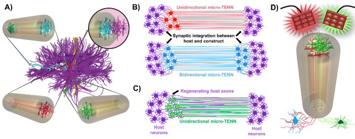
图1
图1:微组织工程神经网络(微型TENN)发展背后的原理和启示。A )Micro-TENN模拟大脑连接体(紫色)的细胞结构,其中功能上不同的区域以单向(红色,绿色)或双向(蓝色)方式通过长对准的轴突束连接。作为一个例子,微型TENN可以重建皮质丘脑和黑质纹状体途径或从内嗅皮质到海马的穿孔途径中的丢失的连接(改编自Struzyna ,2015) 1 。 ( B )单向图l和双向微型TENN(分别为红色和蓝色)与主机电路(紫色)进行突触式积分,以用作病变两端之间的功能继电器。 ( C )单向微-TENN(绿色)的轴突区的示意图,作为用于轴突促进再生主轴突(紫色)朝向与微TENN相互作用的靶标的指导。 ( D )使用光生物活性微TENNS作为神经调节剂的概念图,利用与兴奋性或抑制性神经元的突触整合(底部)。 请点击此处查看此图的较大版本。

目前的手稿详细介绍了使用胚胎衍生的大脑皮质神经元制造微型TENN的方法。值得注意的是,微TENN可以用其他类型的神经细胞制造。例如充足的,成功的微型TENN发展的初步报告是背根神经节(DRG)神经元32 。可以通过将液体琼脂糖加入到定制的激光切割的圆柱形通道阵列或毛细管中(均包含对准的针灸针)来产生水凝胶微柱( 图2A )。针形成内腔并确定微柱的内径(ID),而激光切割装置中的毛细管ID和圆柱体的直径决定了构造体的外径(OD)。 OD和ID可以根据所需的应用选择,分别选择器件/毛细管和针灸针的不同直径。微柱的长度也可以变化;到目前为止,我们已经报道了长达10毫米的微型TENN的建设10 ,并且正在积极追求更长的长度。琼脂糖凝胶和针灸后eedles被去除,通常由I型胶原和层粘连蛋白组成的ECM溶液被添加到构建体的内腔( 图2C )。 ECM核心提供支架,以支持神经细胞粘附和轴突生长。最初,使用解离的细胞悬浮液10,31,32,将原代大鼠皮质神经元电镀在微量柱中。然而,这种方法在所有情况下都没有产生靶细胞结构,其被定义为限于微柱末端的神经细胞体,其中心腔由纯对准的轴突区组成。从那时起,使用强制神经元聚集方法(基于Ungrin 等人改编的协议)使得具有理想结构的微TENN更加可靠和一致的制造( 图2B44 。除了描述当前方法论,本文将展示微型TENNs的代表性相位对比和共聚焦图像,证明随着时间的推移形成轴突束,以及完成的目标细胞结构。该手稿还将扩大协议的重要方面,还将展示微型TENN技术的未来挑战和未来发展方向。
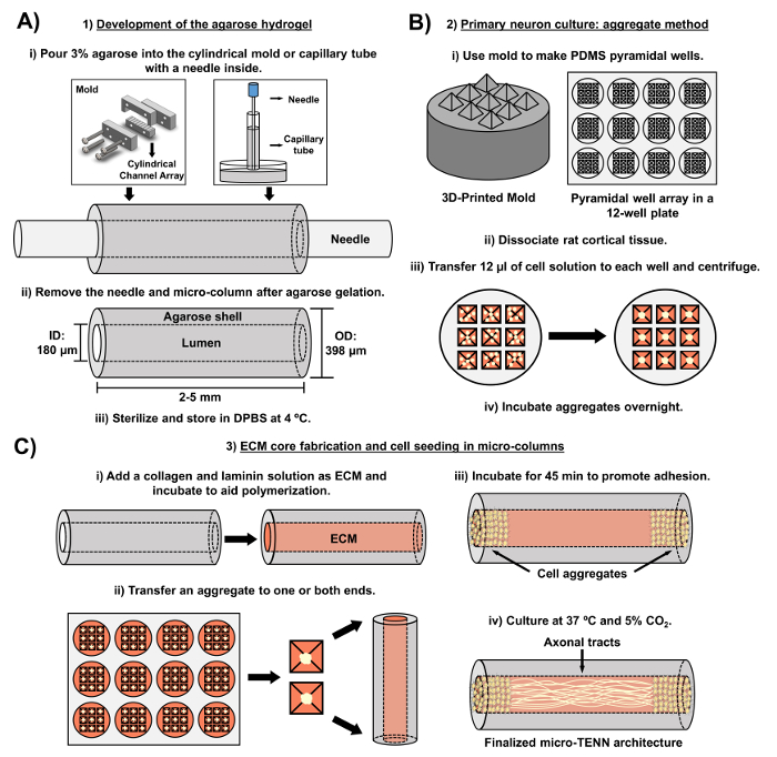
图2
图2:三阶段微型TENN制造工艺的示意图。A )琼脂糖水凝胶的开发:(i)最初,将小针灸针( 例如 ,直径为180-350μm)插入定制的激光切割模具或毛细管的圆柱形通道中( 例如 ,直径380-700μm)。在下一步中,将DPBS中的液体琼脂糖引入圆柱形通道或毛细管中。 (ii)琼脂糖凝胶后,取出针头将模具拆开以产生中空的琼脂糖微柱。 (iii)然后将这些构建体灭菌并储存在DPBS中。 ( B )主要神经元培养和聚集方法:(i)神经元聚集在适合于12孔培养板的孔中的3D印刷模具铸造的金字塔微孔阵列中进行。 (ii)微TENN包括从胚胎-18天大鼠的胎脑分离的原代大鼠神经元。在用胰蛋白酶-EDTA和DNA酶I组织解离之后,制备密度为1.0-2.0×10 6个细胞/ mL的细胞溶液。 (iii)将12μL该溶液转移到锥体微孔阵列中的每个孔中。将含有这些微孔的板离心以产生细胞聚集体。 (iv)然后将它们在电镀之前在微柱中孵育过夜。 ( C )ECM核心制备和细胞接种:(i)细胞播种前,将含有1mg / mL I型胶原和1mg / mL的ECM溶液层粘连蛋白转移到微TENN的内部并使其聚合。 (ii)根据单向或双向微型TENN是否正在制造,聚集体分别放置在微柱的一个或两个极端。 (iii)经过一段时间的温育以促进粘附,将微型TENN在充满补充的胚胎神经元基础培养基的培养皿中培养。 (iv)在培养3-5天后,最终的微TENN结构应在微柱的极端处展示细胞聚集体,其轴突束跨越其长度。 请点击此处查看此图的较大版本。

Protocol

涉及动物的所有手续均由宾夕法尼亚大学的机构动物保护和使用委员会和Michael J. Crescenz退伍军人事务医疗中心批准,并遵守NIH公共卫生服务政策中关于人道关怀和使用实验室的指导原则动物(2015)。 1.开发琼脂糖水凝胶(微TENNs的无细胞成分) 琼脂糖溶液制备 在生物安全柜内,通过将20 mL Dulbecco磷酸缓冲盐水(DPBS)转移到两个10 cm培养皿中的每一…

Representative Results

使用相差显微镜监测微TENNs,以评估其细胞结构和轴突生长( 图4 )。在单向,2mm长的微TENNs中,神经元聚集体被限制在微柱的一端并且通过内核投射一束轴突。轴突将体外的整个长度跨越5天(DIV)( 图4A )。双向2 mm长的微型TENN中的初始轴突生长率较高,因为轴突通过3 DIV跨越整个微柱( 图4B )。通过5 DIV,双向微TENNs特征是?…

Discussion

CNS损伤和疾病通常导致构成脑连接的长途轴突途径的丧失或功能障碍,伴有或不伴有神经元变性。由于中枢神经系统有限的能力来促进神经发生和再生,所以复合化。尽管追求修复策略,如生长因子,细胞和生物材料传递作为个体或组合方法,这些技术不能同时解释神经元细胞的退化和轴突连接的损失14,22。技术上的这些差距限制了以受控和持续的方式修复,改变和探测神经网络的能力。因此,?…

Disclosures

The authors have nothing to disclose.

Acknowledgements

财政支持由美国国立卫生研究院U01-NS094340(Cullen),T32-NS043126(Harris)和F31-NS090746(Katiyar)提供),Michael J.Fox基金会(治疗管道计划#9998(Cullen)), Penn医学神经科学中心试点奖(Cullen),国家科学基金会(研究生研究奖学金DGE-1321851(Struzyna和Adewole)),退伍军人事务部(RR&D优异评估#B1097-I(Cullen)),美国协会的神经外科医生和神经外科医生大会(2015-2016 – 神经损伤与重症监护(Petrov)Codman研究金)和美国陆军医学研究和物资司令部(#W81XWH-13-207004(Cullen)和W81XWH-15-1- 0466(Cullen))。

Materials

Laser cutter Universal Laser Systems PLS4.75 Used to fabricate the laser-cut micro-channel mold.
Laser-cut micro-column fabrication device Custom-made ————– Contact our research group if interested. Dimensions and blueprints provided in the manuscript.
Screws ————– ————– #4-40 with a thread diameter of 3.05 mm
Nuts ————– ————– #4-40 with a thread diameter of 3.05 mm
Acupuncture needle (180 µm diameter) Lhasa Medical sj.16X40 The diameter may be varied according to the desired size for the micro-column lumen.
Petri dish Fisher 08772B
Dulbecco's phosphate buffered saline (DPBS) Invitrogen 14200075
Polystyrene disposable serological pipet Fisher 13-678-11D
Agarose Sigma A9539-50G
Capillary tube (398 µm diameter) Fisher 21170D The diameter may be varied according to the desired size for the micro-column shell.
Hot plate Fisher SP88857200
Magnetic bar Fisher 1451352
Micropipette Sigma Z683884-1EA
25 mm gauge needle Fisher 14-826-49
Microscalpel Roboz Surgical RS-6270
Scissors Fine Science Tools 14081-09
Forceps World Precision Instruments 501985
Hot bead sterilizer Sigma Z378550-1EA
Stereoscope Nikon SMZ800N Used for all dissection steps and for micro-TENN fabrication.
Rat tail type I collagen Corning 354236 Maintain at 4 ºC and remove only when needed.  Use ice to preserve its temperature when in use.
Microcentrifuge tube Fisher 02-681-256
Mouse laminin Corning 354232 Maintain at 4 ºC and remove only when needed.  Use ice to preserve its temperature when in use.
Neurobasal medium Invitrogen 21103049 Basal medium for the culture of pre-natal and embryonic neuronal cells. Store at 4ºC and warm at 37 ºC before use.
Sodium hydroxide (NaOH) Fisher SS2661
Hydrochloric acid (HCl) Fisher SA48-1
Litmus paper Fisher 09-876-18
Hank's balanced salt solution (HBSS) Invitrogen 14170112 Store at 4 ºC.
0.25% Trypsin-EDTA Invitrogen 25200056 Store at -20 ºC and warm at 37 ºC before use.
Bovine pancreatic deoxyribonuclease (DNase) I Sigma 10104159001 Store at -20 ºC and warm at 37 ºC before use.
B-27 Supplement Invitrogen 12587010 Supplement added to Neurobasal medium for the culture of hippocampal and cortical neurons. Store at -20 ºC and warm at 37 ºC before use.
L-glutamine  Invitrogen 35050061 Store at -20 ºC and warm at 37 ºC before use.
Sprague Dawley embryonic day 18 rats Charles River Strain 001
Pasteur pipette Fisher 22-042816
15 mL centrifuge tube EMESCO 1194-352099
Vortex Fisher 02-215-414
Centrifuge Fisher 05-413-115
Hemocytometer Fisher 02-671-6
Objet30 3D-Printer Stratasys  ————– Used to fabricate the pyramidal micro-well molds.
3D-printed pyramidal well mold Custom-made ————– Contact our research group if interested. Dimensions and blueprints provided in the manuscript.
Polydimethylsiloxane (PDMS) and curing agent Fisher NC9285739 Comes as kit with elastomer and curing agent. Use inside a chemical fume hood.
Funnel Fisher 10-348C
1 ml pipette bulb Sigma Z509035
Micro-spatula Fisher S50821
12-well culture plate EMESCO 1194-353043
Oven Fisher 11-475-154
Incubator Fisher 13 998 076
AAV1.Syn.GCaMP6f.WPRE.SV40 UPenn Vector Core 36373 Store at -80ºC. Commercially available adeno-associated virus (AAV) with the GCaMP6f calcium indicator.
Formaldehyde 40% Fisher F77P-4 Formaldehyde is a toxic compound known to be carcinogenic, and must be disposed of in a separate container. 
Triton X-100 Sigma T8787 Non-ionic surfactant used to permeabilize cell membranes.
Horse serum Gibco 16050-122
Mouse anti-Tuj-1/beta-III tubulin primary antibody Sigma T8578-200UL Store at -20ºC.
Rabbit anti-synapsin 1 primary antibody Synaptic Systems 106-001 Store at -20ºC.
Donkey anti-mouse 568 secondary antibody Invitrogen A10037 Store at 4ºC.
Donkey anti-rabbit 488 secondary antibody Invitrogen A21206 Store at 4ºC.
Hoechst 33342, Trihydrochloride Invitrogen H3570 Store at 4ºC.  Hoechst is a known mutagen that should be treated as a carcinogen.  Therefore, it must be disposed of in a separate container.
A1RSI Laser Scanning Confocal Microscope  Nikon ————– Used for taking the confocal reconstructions of immunolabeled constructs.
Eclipse Ti-S Microscope  Nikon ————– Used for taking the phase-contrast images.  With digital image acquisition using a QiClick camera interfaced with Nikon Elements Basic Research software (4.10.01).
High-speed Fluorescence Microscope Nikon ————– Nikon Eclipse Ti microscope paired with an ANDOR Neo/Zyla camera for calcium imaging.
NIS Elements AR 4.50.00 Software Nikon Instruments ————– Used to identify calcium transients from the recordings taken with the high-speed fluorescence microscope. 

References

  1. Struzyna, L. A., Harris, J. P., Katiyar, K. S., Chen, H. I., Cullen, D. K. Restoring nervous system structure and function using tissue engineered living scaffolds. Neural Regen. Res. 10 (5), 679-685 (2015).
  2. Tallantyre, E. C., Bø, L., et al. Clinico-pathological evidence that axonal loss underlies disability in progressive multiple sclerosis. Mult. Scler. 16 (4), 406-411 (2010).
  3. Cheng, H. C., Ulane, C. M., Burke, R. E. Clinical Progression in Parkinson Disease and the Neurobiology of Axons. Ann. Neurol. 67 (6), 715-725 (2010).
  4. Johnson, V. E., Stewart, W., Smith, D. H. Axonal pathology in traumatic brain injury. Exp. Neurol. 246, 35-43 (2013).
  5. Hinman, J. D. The back and forth of axonal injury and repair after stroke. Curr. Opin. Neurol. 27 (6), 615-623 (2014).
  6. Li, X., Katsanevakis, E., Liu, X., Zhang, N., Wen, X. Engineering neural stem cell fates with hydrogel design for central nervous system regeneration. Prog. Polym. Sci. 37 (8), 1105-1129 (2012).
  7. Horner, P. J., Gage, F. H. Regenerating the damaged central nervous system. Nature. 407 (6807), 963-970 (2000).
  8. Yiu, G., He, Z. Glial inhibition of CNS axon regeneration. Nat. Rev. Neurosci. 7 (8), 617-627 (2006).
  9. Montani, L., Petrinovic, M. M. Targeting Axonal Regeneration: The Growth Cone Takes the Lead. J. Neurosci. 34 (13), 4443-4444 (2014).
  10. Struzyna, L. A., Wolf, J. A., et al. Rebuilding Brain Circuitry with Living Micro-Tissue Engineered Neural Networks. Tissue Eng. Part A. 21 (21-22), 2744-2756 (2015).
  11. Huebner, E. a., Strittmatter, S. M. Axon Regeneration in the Peripheral and Central Nervous Systems. Results Probl. Cell Differ. 48, 339-351 (2009).
  12. Benowitz, L. I., Yin, Y. Combinatorial Treatments for Promoting Axon Regeneration in the CNS: Strategies for Overcoming Inhibitory Signals and Activating Neurons’ Intrinsic Growth State. Dev. Neurobiol. 67 (9), 1148-1165 (2007).
  13. Lie, D. C., Song, H., Colamarino, S. A., Ming, G., Gage, F. H. Neurogenesis in the Adult Brain: New Strategies for Central Nervous System Diseases. Annu. Rev. Pharmacol. Toxicol. 44, 399-421 (2004).
  14. Gao, Y., Yang, Z., Li, X. Regeneration strategies after the adult mammalian central nervous system injury-biomaterials. Regen. Biomater. 3 (2), 115-122 (2016).
  15. Benfey, M., Aguayo, A. J. Extensive elongation of axons from rat brain into peripheral nerve grafts. Nature. 296 (11), 150-152 (1982).
  16. David, S., Aguayo, A. J. Axonal Elongation into Peripheral Nervous System “Bridges” after Central Nervous System Injury in Adult Rats. Science. 214 (4523), 931-933 (1981).
  17. Shoichet, M. S., Tate, C. C., Baumann, M. D., LaPlaca, M. C. Strategies for Regeneration and Repair in the Injured Central Nervous System. Indwelling Neural Implant. Strateg. Contend. with Vivo Environ. , (2008).
  18. Lu, P., Tuszynski, M. H. Growth factors and combinatorial therapies for CNS regeneration. Exp. Neurol. 209 (2), 313-320 (2008).
  19. Curinga, G., Smith, G. M. Molecular/genetic manipulation of extrinsic axon guidance factors for CNS repair and regeneration. Exp. Neurol. 209 (2), 333-342 (2008).
  20. Mehta, S. T., Luo, X., Park, K. K., Bixby, J. L., Lemmon, V. P. Hyperactivated Stat3 boosts axon regeneration in the CNS. Exp. Neurol. 280, 115-120 (2016).
  21. Elliott Donaghue, I., Tam, R., Sefton, M. V., Shoichet, M. S. Cell and biomolecule delivery for tissue repair and regeneration in the central nervous system. J. Control. Release. 190, 219-227 (2014).
  22. Tam, R. Y., Fuehrmann, T., Mitrousis, N., Shoichet, M. S. Regenerative therapies for central nervous system diseases: a biomaterials approach. Neuropsychopharmacology. 39 (1), 169-188 (2014).
  23. Cullen, D. K., Wolf, J. A., Smith, D. H., Pfister, B. J. Neural Tissue Engineering for Neuroregeneration and Biohybridized Interface Microsystems In vivo (Part 2). Crit. Rev. Biomed. Eng. 39 (3), 243-262 (2011).
  24. Han, Q., Jin, W., et al. The promotion of neural regeneration in an extreme rat spinal cord injury model using a collagen scaffold containing a collagen binding neuroprotective protein and an EGFR neutralizing antibody. Biomaterials. 31 (35), 9212-9220 (2010).
  25. Suzuki, H., Kanchiku, T., et al. Artificial collagen-filament scaffold promotes axon regeneration and long tract reconstruction in a rat model of spinal cord transection. Med. Mol. Morphol. 48 (4), 214-224 (2015).
  26. Silva, N. A., Salgado, A. J., et al. Development and Characterization of a Novel Hybrid Tissue Engineering-Based Scaffold for Spinal Cord Injury Repair. Tissue Eng. Part A. 16 (1), 45-54 (2009).
  27. Moore, M. J., Friedman, J. A., et al. Multiple-channel scaffolds to promote spinal cord axon regeneration. Biomaterials. 27 (3), 419-429 (2006).
  28. Tsai, E. C., Dalton, P. D., Shoichet, M. S., Tator, C. H. Synthetic hydrogel guidance channels facilitate regeneration of adult rat brainstem motor axons after complete spinal cord transection. J. Neurotrauma. 21 (6), 789-804 (2004).
  29. Chen, B. K., Knight, A. M., et al. Axon regeneration through scaffold into distal spinal cord after transection. J. Neurotrauma. 26 (10), 1759-1771 (2009).
  30. Jain, A., Kim, Y. -. T., McKeon, R. J., Bellamkonda, R. V. In situ gelling hydrogels for conformal repair of spinal cord defects, and local delivery of BDNF after spinal cord injury. Biomaterials. 27 (3), 497-504 (2006).
  31. Harris, J. P., Struzyna, L. A., Murphy, P. L., Adewole, D. O., Kuo, E., Cullen, D. K. Advanced biomaterial strategies to transplant preformed micro-tissue engineered neural networks into the brain. J. Neural Eng. 13 (1), 16019-16037 (2016).
  32. Cullen, D. K., Tang-Schomer, M. D., et al. Microtissue engineered constructs with living axons for targeted nervous system reconstruction. Tissue Eng. Part A. 18 (21-22), 2280-2289 (2012).
  33. Tate, M. C., Shear, D. A., Hoffman, S. W., Stein, D. G., Archer, D. R., LaPlaca, M. C. Fibronectin promotes survival and migration of primary neural stem cells transplanted into the traumatically injured mouse brain. Cell Transplant. 11 (3), 283-295 (2002).
  34. Denham, M., Parish, C. L., et al. Neurons derived from human embryonic stem cells extend long-distance axonal projections through growth along host white matter tracts after intra-cerebral transplantation. Front. Cell. Neurosci. 6 (11), (2012).
  35. Fawcett, J. W., Barker, R. A., Dunnet, S. B. Dopaminergic neuronal survival and the effects of bFGF in explant, three dimensional and monolayer cultures of embryonic rat ventral mesencephalon. Exp. Brain Res. 106 (2), 275-282 (1995).
  36. Mine, Y., Tatarishvili, J., Oki, K., Monni, E., Kokaia, Z., Lindvall, O. Grafted human neural stem cells enhance several steps of endogenous neurogenesis and improve behavioral recovery after middle cerebral artery occlusion in rats. Neurobiol. Dis. 52, 191-203 (2013).
  37. Ren, H., Chen, J., Wang, Y., Zhang, S., Zhang, B. Intracerebral neural stem cell transplantation improved the auditory of mice with presbycusis. Int. J. Clin. Exp. Pathol. 6 (2), 230-241 (2013).
  38. Sinclair, S. R., Fawcett, J. W., Dunnett, S. B. Dopamine cells in nigral grafts differentiate prior to implantation. Eur. J. Neurosci. 11 (12), 4341-4348 (1999).
  39. Tate, C. C., Shear, D. A., Stein, D. G., Tate, M., LaPlaca, M. C., Archer, D. R. Laminin and fibronectin scaffolds enhance neural stem cell transplantation into the injured brain. J. Tissue Eng. Regen. Med. 3 (3), 208-217 (2009).
  40. Yoo, S. J., Kim, J., Lee, C. -. S., Nam, Y. Simple and novel three dimensional neuronal cell culture using a micro mesh scaffold. Exp. Neurobiol. 20 (2), 110-115 (2011).
  41. Chen, H. I., Jgamadze, D., Serruya, M. D., Cullen, D. K., Wolf, J. A., Smith, D. H. Neural Substrate Expansion for the Restoration of Brain Function. Front. Syst. Neurosci. 10, (2016).
  42. Cullen, D. K., Wolf, J. A., Vernekar, V. N., Vukasinovic, J., LaPlaca, M. C. Neural tissue engineering and biohybridized microsystems for neurobiological investigation in vitro (Part 1). Crit. Rev. Biomed. Eng. 39 (3), 201-240 (2011).
  43. Struzyna, L. A., Katiyar, K., Cullen, D. K. Living scaffolds for neuroregeneration. Curr. Opin. Solid State Mater. Sci. 18 (6), 308-318 (2014).
  44. Ungrin, M. D., Joshi, C., Nica, A., Bauwens, C., Zandstra, P. W. Reproducible, ultra high-throughput formation of multicellular organization from single cell suspension-derived human embryonic stem cell aggregates. PLoS One. 3 (2), (2008).
  45. Dahotre, N. B., Harimkar, S. . Laser fabrication and machining of materials. , (2008).
  46. Kutter, J. P., Klank, H., Snakenborg, D. Microstructure fabrication with a CO2 laser system. J. Micromechanics Microengineering. 14 (2), (2004).
  47. Spicar-Mihalic, P., Houghtaling, J., Fu, E., Yager, P., Liang, T., Toley, B. CO2 laser cutting and ablative etching for the fabrication of paper-based devices. J. Micromechanics Microengineering. 23 (6), (2013).
  48. Pacifici, M., Peruzzi, F. Isolation and culture of rat embryonic neural cells: a quick protocol. J. Vis. Exp. (63), (2012).
  49. Cullen, D. K., Gilroy, M. E., Irons, H. R., Laplaca, M. C. Synapse-to-neuron ratio is inversely related to neuronal density in mature neuronal cultures. Brain Res. 1359, 44-55 (2010).
  50. Huang, J. H., Cullen, D. K., et al. Long-Term Survival and Integration of Transplanted Engineered Nervous Tissue Constructs Promotes Peripheral Nerve Regeneration. Tissue Eng. Part A. 15 (7), 1677-1685 (2009).
  51. Katiyar, K. S., Winter, C. C., Struzyna, L., Harris, J. P., Cullen, D. K. Mechanical elongation of astrocyte processes to create living scaffolds for nervous system regeneration. J. Tissue Eng. Regan. Med. , (2016).
  52. Howard, M. A., Baraban, S. C. Synaptic integration of transplanted interneuron progenitor cells into native cortical networks. J. Neurophysiol. 116 (2), 472-478 (2016).
  53. Wernig, M., Benninger, F., et al. Functional Integration of Embryonic Stem Cell-Derived Neurons In Vivo. J. Neurosci. 24 (22), 5258-5268 (2004).
  54. Ganguly, K., Poo, M. Activity-dependent neural plasticity from bench to bedside. Neuron. 80 (3), 729-741 (2013).
  55. Dancause, N. Extensive Cortical Rewiring after Brain Injury. J. Neurosci. 25 (44), 10167-10179 (2005).
  56. Winter, C. C., Katiyar, K. S., et al. Transplantable living scaffolds comprised of micro-tissue engineered aligned astrocyte networks to facilitate central nervous system regeneration. Acta Biomater. 38, 44-58 (2016).
  57. Adewole, D. O., Serruya, M. D., et al. The Evolution of Neuroprosthetic Interfaces. Crit. Rev. Biomed. Eng. 44 (1-2), 123-152 (2016).
  58. Cullen, D. K., Patel, A., Doorish, J. F., Smith, D. H., Pfister, B. J. Developing a tissue-engineered neural-electrical relay using encapsulated neuronal constructs on conducting polymer fibers. J. Neural Eng. 5 (4), 374-384 (2008).
  59. Cullen, D. K., Stabenfeldt, S. E., Simon, C. M., Tate, C. C., LaPlaca, M. C. In Vitro Neural Injury Model for Optimization of Tissue-Engineered Constructs. J. Neurosci. Res. 85, 3642-3651 (2007).
  60. Irons, H. R., Cullen, D. K., Shapiro, N. P., Lambert, N. A., Lee, R. H., Laplaca, M. C. Three-dimensional neural constructs: a novel platform for neurophysiological investigation. J. Neural Eng. 5 (3), 333-341 (2008).
  61. Morrison, B. I., Cullen, D. K., LaPlaca, M. In Vitro Models for Biomechanical Studies of Neural Tissues. Stud. Mechanobiol. Tissue Eng. Biomater. 3, 247-285 (2011).
  62. Vukasinovic, J., Cullen, D. K., Laplaca, M. C., Glezer, A. A microperfused incubator for tissue mimetic 3D cultures. Biomed. Microdevices. 11 (6), 1155-1165 (2009).
  63. Cullen, D. K., Lessing, M. C., Laplaca, M. C. Collagen-dependent neurite outgrowth and response to dynamic deformation in three-dimensional neuronal cultures. Ann. Biomed. Eng. 35 (5), 835-846 (2007).
  64. LaPlaca, M. C., Vernekar, V. N., Shoemaker, J. T., Cullen, D. K. Three-dimensional neuronal cultures. Methods Bioeng. 3D Tissue Eng. , (2010).
  65. Chwalek, K., Tang-Schomer, M. D., Omenetto, F. G., Kaplan, D. L. In vitro bioengineered model of cortical brain. Nat. Protoc. 10 (9), 1362-1373 (2015).
check_url/55609?article_type=t

Play Video

Cite This Article
Struzyna, L. A., Adewole, D. O., Gordián-Vélez, W. J., Grovola, M. R., Burrell, J. C., Katiyar, K. S., Petrov, D., Harris, J. P., Cullen, D. K. Anatomically Inspired Three-dimensional Micro-tissue Engineered Neural Networks for Nervous System Reconstruction, Modulation, and Modeling. J. Vis. Exp. (123), e55609, doi:10.3791/55609 (2017).

View Video