Summary

신경 시스템 재구성, 변조 및 모델링을 위해 해부학 적으로 영감을받은 3 차원 미세 조직 공학 신경 회로망

Published: May 31, 2017
doi:

Summary

이 원고는 마이크로 조직 공학 신경 네트워크의 제작에 대해 자세히 설명합니다. 3 차원 크기의 마이크론 크기의 구조물은 관 모양의 하이드로 겔에 둘러싸인 집적 된 신경 세포군에 걸친 길쭉한 정렬 축색관으로 구성됩니다. 이러한 살아있는 비계는 신경 회로를 재구성하거나 조절하기위한 기능적 중계 역할을하거나 회색 – 흰색 물질 신경 조직학을 모방 한 생물 여과 테스트 베드 (bifidelic test-bed) 역할을 할 수 있습니다.

Abstract

기능적 회복은 중추 신경계 (CNS) 내의 손상 또는 질병 유발 성 퇴행 후 억제 환경 및 신경 발생에 대한 제한된 용량으로 인해 드물게 발생한다. 우리는 손상된 CNS 내에서 신경 및 축삭 경로 손실을 동시에 처리 할 수있는 전략을 개발 중입니다. 이 원고는 마이크로 조직 공학 신경 네트워크 (micro-TENNs), 뉴런으로 구성된 이식 가능한 구조물 및 센티미터 (centimeters)를 확장 할 수있는 수백 마이크론의 직경을 가진 예비 형성된 하이드로 겔 실린더의 세포 외 매트릭스 (extracellular matrix, ECM) 길이. 신경원 집합체는 3 차원 인장의 극단으로 구분되며 축삭 투영으로 확장됩니다. Micro-TENNs는 CNS 재건을위한 전략으로 두뇌 connectome 세포 구조의 측면을 모방하고 잠재적으로 네트워크 대체 수단을 제공 할 수있는 유일한 자세를 취하고 있습니다. 노이ronal aggregates는 숙주 조직과 시냅스하여 누락되거나 손상된 회로를 복원 및 / 또는 조절하는 새로운 기능적 중계를 형성 할 수있다. 이러한 구조는 또한 세포 재생 및 축삭 경로 찾기를위한 발달 기작을 이용할 수있는 친 재생성 "살아있는 스캐 폴드"로 작용하여 재생 상태를 기반으로 시너지 구조 및 가용성 단서를 제공 할 수 있습니다. Micro-TENN은 액체 하이드로 겔을 종 방향 중심의 바늘이 들어있는 원통형 틀에 주입하여 제조됩니다. 일단 하이드로 겔이 겔화되면 바늘이 제거되어 중공 형 마이크로 컬럼이 남습니다. 루멘에 ECM 솔루션을 추가하여 신경 부착 및 축색 파 성장에 적합한 환경을 제공합니다. 해리 된 뉴런은 미세 기둥의 한쪽 또는 양쪽 끝 부분에 정확한 파종을 위해 기계적으로 응집됩니다. 이 방법론은 장기적으로 돌출 된 축삭 모양의 좁은 구조물을 안정적으로 생산하여 뇌 신경 조직의 특징을 재현 할 수 있습니다. 시냅스 렘unlabelling 및 유 전적으로 코딩 된 칼슘 지시약은 마이크로 TENN이 광범위한 시냅스 분포와 본질적인 전기적 활동을 가지고 있음을 암시합니다. 결과적으로 micro-TENN은 뇌 경로의 신경 외과 재 구축을위한 유망한 전략이며 , 생체 내에서 신경 생리 현상을 연구하기위한 biofidelic 모델로도 적용될 수 있습니다.

Introduction

외상성 뇌 손상 (TBI), 척수 손상 (SCI), 뇌졸중, 알츠하이머 병 및 파킨슨 병과 같은 중추 신경계 (CNS)의 장애 및 질병의 일반적인 특징은 축삭 경로 및 신경 세포의 단절이다 손실 1 , 2 , 3 , 4 , 5 , 6 . 예를 들어, 허혈성 뇌졸중이 치료되지 않으면 축삭이 분당 7 마일의 속도로 손실된다는 것이 추정됩니다. 미국에서만 약 170 만 명의 사람들이 매년 경험하는 TBI의 경우, 초기 손상이 장기간 신경 퇴행성 상태를 유발하기 때문에 축삭 변성이 외상 후 수년 후에도 계속 발생할 수 있습니다 4 . 이러한 유해한 영향을 악화시키는 중추 신경계는 심각한 카파중생을위한 도시 1 , 7 , 8 , 9 . 손상 후, 먼 표적에 대한 유도 지침의 부족, 신경 돌기 성장을 방해하는 미엘린 – 관련 억제제의 존재 및 반응성 성상 교세포 8 , 10 , 11 , 12 에 의한 신경 교뇌 흉터의 형성을 특징으로하는 억제 환경이 발병한다. glial 흉터는 chondroitin sulfate proteoglycans와 같은 분자가 축삭 돌기를 방해하는 생화학 적 및 물리적 장벽으로 작용합니다 8 , 11 . 또한 신경 줄기 세포가 성인 CNS에서 발견 되더라도 신경 발생의 일관된 증거가 후각 구, 해마에서만 발견되어 새로운 신경 세포의 생성이 제한적이다입방 영역, 뇌실 주위 및 척수의 중심 도관 13 , 14 . 이러한 장애물은 상해 또는 질병에 따른 손실 된 뉴런 및 백질 구조의 기능적 회복을 방해하여 종종 이러한 조건의 삶을 변화시키고 연장 된 결과를 초래합니다.

성인 CNS의 재생 능력이 부족 함에도 불구하고 적절한 환경 신호가 호스트 뉴런 15 , 16 , 17 , 18에 제공 되면 축삭 재생이 가능하다는 것이 입증되었습니다. 연구자들은 성장 인자 ( 예 : 신경 성장 인자, 표피 성장 인자, glial 의존 성장 인자 및 신경 영양 인자 3) 및 가소성 및 축색 재생을 자극하는 다른 유도 분자를 전달 및 조작하려고 시도했다 14 ,18 , 19 . 비록 이러한 연구가 성체 축삭 돌기가 성장 인자에 반응 할 수 있다는 것을 확인했지만, 이러한 전략은 혈액 뇌 장벽의 낮은 침투력과 재생을 촉진시키는 데 필요한 특정 공간적, 시간적 변화에 의해 제한된다 14 , 18 , 19 . 다른 접근법은 중추 신경계 뉴런에서 재생 관련 전사 인자의과 활성화에 의존해왔다. 예를 들어, Stat3 전사 인자의 과발현은 시신경에서 축삭 재생을 자극했다. 그럼에도 불구하고, 전사 인자의 생체 분자 전달 및 과발현은 손실 된 연결 집단을 대체하지 못한다. 세포 기반 전략은 주로 신경 줄기 세포 (NSC)를 중추 신경계에 이식하는 것에 중점을두고 있으며, CNS 뉴런을 대체 할 수있는 능력을 활용하고 영양 계수를 방출하며,부상 후 발생하는 신경 발생에 대한 시도를지지한다. 그럼에도 불구하고 이식 된 신경 세포가 생존하고 숙주와 통합되며 상처 부위 6 , 14 , 17 , 21에 공간적으로 제한된 채로 남아있는 방해받는 능력을 포함하여이 접근법을 방해하는 도전 과제가 여전히 남아 있습니다. 또한, 세포 전달만으로 손상되거나 손상된 축삭 경로의 세포 구조를 복원 할 수 없다. 세포 및 약물 / 화학 물질 전달 전략이 직면 한 문제를 해결할 수있는 대체 방법은 이러한 접근법을 생체 물질 14 , 22 , 23 의 사용과 결합하는 것입니다. 하이드로 겔과 같은 생체 재료는 세포 외 매트릭스 (ECM)의 생화학 적 및 물리적 특성을 에뮬레이션 할 수 있으며, 세포 전달을 돕습니다.손상 부위 내에서의 유지, 방출 조절이 가능한 성장 인자 및 기타 생체 활성 물질 전달 22 . 이러한 생체 재료 기반 전략의 매력적인 특성은 발병 지역 24 , 25 , 26 , 27 , 28 , 29 , 30 에 비계를 이식 한 후 생체 내 축삭 재생의 증거를 가져 왔습니다. 그러나, 무 세포 생체 재료 전략은 잃어버린 연결 인구를 대체하지 않는다; 신경 세포, 신경 교세포 또는 신경 전구체 세포의 전달 비히클로 사용될 때, 생체 재료는 장거리 축색 망을 재구성 할 수 없다. CNS 손상 및 질병과 관련된 축삭 경로의 퇴행 및 신경 세포 손실 모두를 다루는 접근법을 개발하는 과제는 여전히 <sup class = "xref"> 31.

우리 연구 그룹은 이전에 아가로 오스 하이드로 겔 -ECM 마이크로 칼럼의 한쪽 또는 양쪽 끝으로 제한되는 신경 세포 몸체로 구성된 "살아있는 비계 (scaffold)"형태의 이식 가능한 미세 조직 공학 신경 네트워크 (마이크로 TENN)의 개발을보고했다 이 3 차원 (3D) 수용체 1 , 10 , 31 , 32 의 내부 전체에 걸쳐 정렬 된 축색 영역이있다. 이 기법과 이전의 접근법의 가장 큰 차이점 중 하나는 micro-TENNs 의 세포 구조 가 in vitro에서 완전히 생성 되어 이후 33 , 34 , 35 , 36 , 37 , 38 ,sup class = "xref"> 39 , 40 , 41 . In vitro 제작은 세포 표현형 및 배향, 기계적 / 물리적 특성, 생화학 적 신호 및 외인성 인자에 대한 광범위한 공간 및 시간 제어 기능을 제공하여 임플란트 후 이러한 인공 지지체를 숙주와 통합하는 데 효과적입니다. Micro-TENN은 해부학 적으로 영감을받습니다. 뇌의 신경 기능을 모방하기 때문에 뇌의 다른 기능 영역을 연결하는 것과 유사한 축삭 영역을 보이기 때문입니다 ( 그림 1A ) 1 . 따라서이 전략은 병변 부위에 이식 한 후 잃어버린 백질 물질과 뉴런을 물리적으로 대체 할 수 있습니다. 이 기술은 또한 방사상의 glial 세포와 개척 축삭에 의해 형성된 "natural living scaffolds"가 세포의 길 찾기 가이드 역할을하는 발달 기작으로부터 영감을 얻습니다subventricular zone과 axonal outgrowth에서의 이동 43 . 이러한 메커니즘은 신경 세포 이동 및 축삭 – 중재 축삭 돌기 ( 그림 1C ) 43에 의한 축삭 재생을위한 살아있는 경로를 제시 할 수있는 마이크로 TENNs의 정렬 axonal 영역에서 recapitulated 있습니다. 또한이 전략은 마이크로 TENN 뉴런과 기본 회로 사이의 시냅스 통합을 활용하여 기능적 복구 ( 그림 1B ) 43 에 기여할 수있는 새로운 릴레이를 형성합니다. 시냅스 형성 용량은 또한이 접근법에 네트워크 피드백에 따라 CNS를 조절하고 숙주 조직에 반응하는 능력을 부여 할 수있다. 예를 들어, 살아있는 발판의 optogenetically 활성 뉴런은 시냅스 상호 작용을 통해 호스트 뉴런을 변조 자극 수 있습니다 ( 그림 1D ).

또한, 생체 재료 기반 관상 면상 구마이크로 TENN의 작용은 세포 부착, 성장, 신경 돌기 확장 및 신호 전달을위한 적절한 환경을 제공하는 반면, 구조물의 미니어처 차원은 잠재적으로 최소 침습성 이식을 허용하고 부분적으로 격리 된 미세 환경을 제공하여 뇌에 점진적으로 통합시킵니다. 사실, 최근의 연구 결과는 쥐의 뇌에 이식 된 이후에 신경 경로를 모방 할 수있는 마이크로 TENN의 잠재력을 보여주었습니다. stereotaxic microinjection 후, 우리는 이전에 마이크로 TENN의 연결의 생존, axonal tract 건축의 유지 및 생체 내 에서 적어도 1 개월까지의 숙주 피질로의 신경 돌기 연장을보고했다. 또한, synapsin으로 라벨링은 원시 조직과의 시냅스 통합의 조직 학적 증거를 제공했다. 전반적으로 마이크로 TENN은 손상된 부분을 재구성하고 변조하는 데 적합 할 수 있습니다CNS는 잃어버린 뉴런을 교체하고, 숙주 회로와 시냅스 적으로 통합하고, 축삭 세포 구조를 잃어버린 채로 복원하며, 경우에 따라 축삭 재생 축삭에 적절한 길 찾기 신호를 제공함으로써 가능합니다.

그림 1
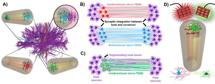
그림 1 : 마이크로 조직 공학 신경 네트워크 (마이크로 TENN) 개발의 원리와 영감 ( A ) Micro-TENN은 기능적으로 구별되는 영역이 단방향 (적색, 녹색) 또는 양방향 (파란색) 방식으로 길고 정렬 된 축색 영역에 의해 연결되는 brain connectome (자주색)의 세포 구조를 모방합니다. 예를 들어, micro-TENNs는 대뇌 피 질 및 흑질 선 경로에서, 또는 entorhinal cortex에서 해마에 이르는 경로에서 분실 된 연결을 재구성 할 수있다 (Struzyna et al. , 2015). ( B ) 단방향의 다이어그램l 및 양방향 micro-TENN (각각 빨간색과 파란색)을 사용하여 병변의 양쪽 끝 사이의 기능 중계 역할을하는 호스트 회로 (자주색)와 통합됩니다. ( C ) micro-TENN이 상호 작용하는 표적을 향한 축색 돌기 (보라색)의 축삭 촉진 재생을위한 가이드 역할을하는 단 향성 마이크로 TENN (녹색)의 축삭 덩어리의 개략도. ( D ) 흥분성 또는 억제 성 뉴런과의 시냅스 통합을 이용하여 신경 조절기로서의 광학 유전 학적으로 활성 인 마이크로 TENNS의 사용에 대한 개념도. 이 그림의 더 큰 버전을 보려면 여기를 클릭하십시오.

현재의 원고는 배아 적으로 추출 된 대뇌 피질 뉴런을 사용하여 마이크로 TENN을 제조하는 데 사용 된 방법론을 상세히 설명합니다. 주목할 만하게, micro-TENNs는 다른 종류의 신경 세포로 만들어 질 수있다. 예를 들어충분한, 마이크로 TENN 개발 성공의 초기 보고서는 지느러미 뿌리 신경절 (DRG) 뉴런을 특징으로합니다 32 . 하이드로 겔 미세 기둥은 주문 제작 된 레이저 컷 원통형 채널 어레이 또는 모세 혈관 튜브에 액체 아가로 오스를 추가하여 생성 할 수 있습니다 ( 그림 2A ). 바늘은 내강을 형성하고 미세 기둥의 내경 (ID)을 결정하는 반면 모세관 ID 및 레이저 절단 장치의 기둥 직경은 구조체의 외경 (OD)을 결정합니다. OD 및 ID는 장치 / 모세관 및 침침 바늘에 각각 다른 직경을 선택하여 원하는 용도에 따라 선택할 수 있습니다. 미세 기둥의 길이 또한 다양 할 수 있습니다. 지금까지 우리는 길이가 20mm 인 마이크로 TENN의 건설을보고했으며, 더 긴 길이를 적극적으로 추구하고 있습니다. 아가로 오스 겔과 침술 후Eedles를 제거하면 일반적으로 I 형 콜라겐과 라미닌으로 구성된 ECM 용액을 구조물의 루멘에 첨가합니다 ( 그림 2C ). ECM 코어는 신경 세포 접착 및 축색 돌기를지지하기위한 지지체를 제공한다. 초기에, 일차 쥐 대뇌 피질 뉴런은 해리 된 세포 현탁액 10 , 31 , 32 를 사용하여 마이크로 칼럼에 도금되었다. 그러나,이 접근법은 모든 경우에 표적 세포 구조를 생성하지 않았는데, 이는 뉴 칼럼의 말단에 한정된 신경 세포 몸체로 정의되었고, 중심 루멘은 순수한 정렬 된 축삭 영역으로 구성되었다. 그 이후 강제적 인 신경 집계 방법 (Ungrin et al .의 프로토콜을 기반으로 함)을 사용하여 이상적인 구조 ( 그림 2B )로 마이크로 TENN을보다 안정적이고 일관되게 제작할 수있었습니다. 전류를 설명하는 것 외에도방법론에서,이 기사는 시간이 지남에 따라 axonal tracts의 형성을 증명하는 micro-TENN의 대표적인 위상 – 대조 및 공 촛점 이미지뿐만 아니라 최종 표적 세포 구조를 보여줄 것이다. 이 원고는 프로토콜의 중요한 측면과 마이크로 TENN 기술의 향후 과제 및 향후 방향으로 확장 될 것입니다.

그림 2
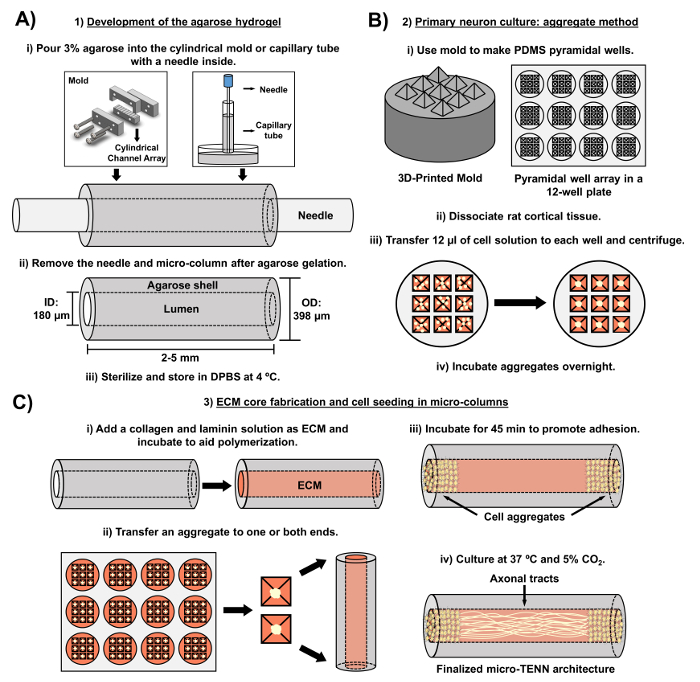
그림 2 : 3 단계 마이크로 TENN 제조 공정의 개략도. ( A ) 아가로 오스 하이드로 겔의 개발 : (i) 처음에는 직경이 180-350 μm 인 작은 침침이 맞춤형 레이저 컷 몰드 또는 모세관 튜브의 원통형 채널에 삽입된다. , 직경 380-700 μm). 다음 단계에서, DPBS의 액상 아가로 오스가 원통형 채널 또는 모세관에 도입된다. (ii) 아가 로스 젤 후, 바늘을 제거하고주형을 분해하여 중공 아가 로스 미세 기둥을 수득한다. (iii) 이들 구조체는 DPBS에 살균되고 저장된다. ( B ) 일차 뉴런 문화와 집계 방법 : (1) 뉴런 집합은 12 잘 배양 접시의 우물에 맞는 3D 인쇄 금형에서 캐스팅 피라미드 형 마이크로 우물 어레이에서 수행됩니다. (ii) Micro-TENN은 배아 일 -18 쥐의 태아 두뇌에서 해리 된 일차 쥐 뉴런을 포함한다. 트립신 -EDTA 및 DNase I로 조직 해리시킨 후, 1.0-2.0 x 10⁶ 세포 / mL의 밀도를 갖는 세포 용액을 제조 하였다. (iii)이 용액 12 μL를 피라미드 마이크로 웰 어레이의 각 웰로 옮긴다. 이러한 마이크로 우물을 포함하는 플레이트를 원심 분리하여 세포 응집체를 생성한다. (iv) 이들을 마이크로 칼럼에서 도금하기 전에 밤새 배양한다. ( C ) ECM 코어 제작 및 세포 파종 : (i) 세포 파종 전에, 1 mg / mL 타입 I 콜라겐 및 1 mg / mL를 함유하는 ECM 용액라미닌은 마이크로 TENN의 내부로 전달되어 중합 될 수있다. (ii) 단방향 또는 양방향 micro-TENN이 제조되는지 여부에 따라, 응집체는 마이크로 컬럼의 한쪽 또는 양쪽 극단에 각각 위치한다. (iii) 접착을 촉진하기위한 배양 기간 후, micro-TENNs를 보충 된 배아 뉴론 기저 배지로 침수시킨 페트리 접시에서 배양한다. (iv) 배양 3 ~ 5 일 후, 최종 마이크로 -TENN 구조는 마이크로 – 칼럼의 극단에서 세포 집합체를 보여야하며, 길이에 걸친 축삭 영역이 있어야한다. 이 그림의 더 큰 버전을 보려면 여기를 클릭하십시오.

Protocol

동물을 포함한 모든 절차는 펜실베이니아 대학의 기관 동물 관리 및 사용위원회와 마이클 J. 크레센츠 재향 군인 의료 센터 (American J. Crescenz Veterans Affairs Medical Center)의 승인을 얻었으며 NIH 공중 보건 서비스 정책에 규정 된 지침에 따라 실험실의 인간애 관리 및 사용 동물 (2015). 1. 아가로 오스 하이드로 겔 (마이크로 TENN의 무 세포 성분) 아가로 오스 용액 준비</…

Representative Results

Micro-TENN은 위상차 현미경을 사용하여 모니터하여 세포 구조 및 축색 돌기를 평가했습니다 ( 그림 4 ). 단방향, 2mm 길이의 마이크로 -TENN 내에서, 뉴런 집합체는 마이크로 – 컬럼의 한쪽 말단으로 제한되고 내부 코어를 통해 축색 돌기가 투영되었다. Axons 은 체외에서 5 일 (DIV) ( 그림 4A ) 열의 전체 길이를 스팬. 축색 돌기가 3 DIV ( 그림 4B<…

Discussion

CNS 손상 및 질병은 전형적으로 부수적 인 신경 세포 변성의 유무에 관계없이 뇌 결합체를 구성하는 장거리 축삭 경로의 손실 또는 기능 부전을 야기한다. 이것은 신경 발생 및 재생을 촉진하는 CNS의 제한된 용량에 의해 합성됩니다. 성장 인자, 세포 및 생체 물질 전달과 같은 수복 전략을 개별적 또는 조합 적 접근으로 추구 함에도 불구하고 이러한 기술은 신경 세포의 퇴행과 축삭 연결의 손실을 …

Disclosures

The authors have nothing to disclose.

Acknowledgements

재정 지원은 U01-NS094340 (컬린), T32-NS043126 (해리스), F31-NS090746 (카티 야), 마이클 J. 폭스 재단 (치료 파이프 라인 프로그램 # 9998 (컬린) (Cullen), 미국 과학 재단 (대학원 연구 동창 DGE-1321851 (Struzyna and Adewole)), Veterans Affairs (RR & D Merit Review # B1097-I (Cullen)), Penn Medicine 신경 과학 센터 파일럿 상 신경 외과 의사 및 신경 외과 의사 회의 (2015-2016 년 신경 외상 및 중환자 실에서의 코닥 개인 연구 (Petrov)) 및 미 육군 의학 연구 및 물질 명령 (# W81XWH-13-207004 (컬른) 및 W81XWH- 0466 (컬른)).

Materials

Laser cutter Universal Laser Systems PLS4.75 Used to fabricate the laser-cut micro-channel mold.
Laser-cut micro-column fabrication device Custom-made ————– Contact our research group if interested. Dimensions and blueprints provided in the manuscript.
Screws ————– ————– #4-40 with a thread diameter of 3.05 mm
Nuts ————– ————– #4-40 with a thread diameter of 3.05 mm
Acupuncture needle (180 µm diameter) Lhasa Medical sj.16X40 The diameter may be varied according to the desired size for the micro-column lumen.
Petri dish Fisher 08772B
Dulbecco's phosphate buffered saline (DPBS) Invitrogen 14200075
Polystyrene disposable serological pipet Fisher 13-678-11D
Agarose Sigma A9539-50G
Capillary tube (398 µm diameter) Fisher 21170D The diameter may be varied according to the desired size for the micro-column shell.
Hot plate Fisher SP88857200
Magnetic bar Fisher 1451352
Micropipette Sigma Z683884-1EA
25 mm gauge needle Fisher 14-826-49
Microscalpel Roboz Surgical RS-6270
Scissors Fine Science Tools 14081-09
Forceps World Precision Instruments 501985
Hot bead sterilizer Sigma Z378550-1EA
Stereoscope Nikon SMZ800N Used for all dissection steps and for micro-TENN fabrication.
Rat tail type I collagen Corning 354236 Maintain at 4 ºC and remove only when needed.  Use ice to preserve its temperature when in use.
Microcentrifuge tube Fisher 02-681-256
Mouse laminin Corning 354232 Maintain at 4 ºC and remove only when needed.  Use ice to preserve its temperature when in use.
Neurobasal medium Invitrogen 21103049 Basal medium for the culture of pre-natal and embryonic neuronal cells. Store at 4ºC and warm at 37 ºC before use.
Sodium hydroxide (NaOH) Fisher SS2661
Hydrochloric acid (HCl) Fisher SA48-1
Litmus paper Fisher 09-876-18
Hank's balanced salt solution (HBSS) Invitrogen 14170112 Store at 4 ºC.
0.25% Trypsin-EDTA Invitrogen 25200056 Store at -20 ºC and warm at 37 ºC before use.
Bovine pancreatic deoxyribonuclease (DNase) I Sigma 10104159001 Store at -20 ºC and warm at 37 ºC before use.
B-27 Supplement Invitrogen 12587010 Supplement added to Neurobasal medium for the culture of hippocampal and cortical neurons. Store at -20 ºC and warm at 37 ºC before use.
L-glutamine  Invitrogen 35050061 Store at -20 ºC and warm at 37 ºC before use.
Sprague Dawley embryonic day 18 rats Charles River Strain 001
Pasteur pipette Fisher 22-042816
15 mL centrifuge tube EMESCO 1194-352099
Vortex Fisher 02-215-414
Centrifuge Fisher 05-413-115
Hemocytometer Fisher 02-671-6
Objet30 3D-Printer Stratasys  ————– Used to fabricate the pyramidal micro-well molds.
3D-printed pyramidal well mold Custom-made ————– Contact our research group if interested. Dimensions and blueprints provided in the manuscript.
Polydimethylsiloxane (PDMS) and curing agent Fisher NC9285739 Comes as kit with elastomer and curing agent. Use inside a chemical fume hood.
Funnel Fisher 10-348C
1 ml pipette bulb Sigma Z509035
Micro-spatula Fisher S50821
12-well culture plate EMESCO 1194-353043
Oven Fisher 11-475-154
Incubator Fisher 13 998 076
AAV1.Syn.GCaMP6f.WPRE.SV40 UPenn Vector Core 36373 Store at -80ºC. Commercially available adeno-associated virus (AAV) with the GCaMP6f calcium indicator.
Formaldehyde 40% Fisher F77P-4 Formaldehyde is a toxic compound known to be carcinogenic, and must be disposed of in a separate container. 
Triton X-100 Sigma T8787 Non-ionic surfactant used to permeabilize cell membranes.
Horse serum Gibco 16050-122
Mouse anti-Tuj-1/beta-III tubulin primary antibody Sigma T8578-200UL Store at -20ºC.
Rabbit anti-synapsin 1 primary antibody Synaptic Systems 106-001 Store at -20ºC.
Donkey anti-mouse 568 secondary antibody Invitrogen A10037 Store at 4ºC.
Donkey anti-rabbit 488 secondary antibody Invitrogen A21206 Store at 4ºC.
Hoechst 33342, Trihydrochloride Invitrogen H3570 Store at 4ºC.  Hoechst is a known mutagen that should be treated as a carcinogen.  Therefore, it must be disposed of in a separate container.
A1RSI Laser Scanning Confocal Microscope  Nikon ————– Used for taking the confocal reconstructions of immunolabeled constructs.
Eclipse Ti-S Microscope  Nikon ————– Used for taking the phase-contrast images.  With digital image acquisition using a QiClick camera interfaced with Nikon Elements Basic Research software (4.10.01).
High-speed Fluorescence Microscope Nikon ————– Nikon Eclipse Ti microscope paired with an ANDOR Neo/Zyla camera for calcium imaging.
NIS Elements AR 4.50.00 Software Nikon Instruments ————– Used to identify calcium transients from the recordings taken with the high-speed fluorescence microscope. 

References

  1. Struzyna, L. A., Harris, J. P., Katiyar, K. S., Chen, H. I., Cullen, D. K. Restoring nervous system structure and function using tissue engineered living scaffolds. Neural Regen. Res. 10 (5), 679-685 (2015).
  2. Tallantyre, E. C., Bø, L., et al. Clinico-pathological evidence that axonal loss underlies disability in progressive multiple sclerosis. Mult. Scler. 16 (4), 406-411 (2010).
  3. Cheng, H. C., Ulane, C. M., Burke, R. E. Clinical Progression in Parkinson Disease and the Neurobiology of Axons. Ann. Neurol. 67 (6), 715-725 (2010).
  4. Johnson, V. E., Stewart, W., Smith, D. H. Axonal pathology in traumatic brain injury. Exp. Neurol. 246, 35-43 (2013).
  5. Hinman, J. D. The back and forth of axonal injury and repair after stroke. Curr. Opin. Neurol. 27 (6), 615-623 (2014).
  6. Li, X., Katsanevakis, E., Liu, X., Zhang, N., Wen, X. Engineering neural stem cell fates with hydrogel design for central nervous system regeneration. Prog. Polym. Sci. 37 (8), 1105-1129 (2012).
  7. Horner, P. J., Gage, F. H. Regenerating the damaged central nervous system. Nature. 407 (6807), 963-970 (2000).
  8. Yiu, G., He, Z. Glial inhibition of CNS axon regeneration. Nat. Rev. Neurosci. 7 (8), 617-627 (2006).
  9. Montani, L., Petrinovic, M. M. Targeting Axonal Regeneration: The Growth Cone Takes the Lead. J. Neurosci. 34 (13), 4443-4444 (2014).
  10. Struzyna, L. A., Wolf, J. A., et al. Rebuilding Brain Circuitry with Living Micro-Tissue Engineered Neural Networks. Tissue Eng. Part A. 21 (21-22), 2744-2756 (2015).
  11. Huebner, E. a., Strittmatter, S. M. Axon Regeneration in the Peripheral and Central Nervous Systems. Results Probl. Cell Differ. 48, 339-351 (2009).
  12. Benowitz, L. I., Yin, Y. Combinatorial Treatments for Promoting Axon Regeneration in the CNS: Strategies for Overcoming Inhibitory Signals and Activating Neurons’ Intrinsic Growth State. Dev. Neurobiol. 67 (9), 1148-1165 (2007).
  13. Lie, D. C., Song, H., Colamarino, S. A., Ming, G., Gage, F. H. Neurogenesis in the Adult Brain: New Strategies for Central Nervous System Diseases. Annu. Rev. Pharmacol. Toxicol. 44, 399-421 (2004).
  14. Gao, Y., Yang, Z., Li, X. Regeneration strategies after the adult mammalian central nervous system injury-biomaterials. Regen. Biomater. 3 (2), 115-122 (2016).
  15. Benfey, M., Aguayo, A. J. Extensive elongation of axons from rat brain into peripheral nerve grafts. Nature. 296 (11), 150-152 (1982).
  16. David, S., Aguayo, A. J. Axonal Elongation into Peripheral Nervous System “Bridges” after Central Nervous System Injury in Adult Rats. Science. 214 (4523), 931-933 (1981).
  17. Shoichet, M. S., Tate, C. C., Baumann, M. D., LaPlaca, M. C. Strategies for Regeneration and Repair in the Injured Central Nervous System. Indwelling Neural Implant. Strateg. Contend. with Vivo Environ. , (2008).
  18. Lu, P., Tuszynski, M. H. Growth factors and combinatorial therapies for CNS regeneration. Exp. Neurol. 209 (2), 313-320 (2008).
  19. Curinga, G., Smith, G. M. Molecular/genetic manipulation of extrinsic axon guidance factors for CNS repair and regeneration. Exp. Neurol. 209 (2), 333-342 (2008).
  20. Mehta, S. T., Luo, X., Park, K. K., Bixby, J. L., Lemmon, V. P. Hyperactivated Stat3 boosts axon regeneration in the CNS. Exp. Neurol. 280, 115-120 (2016).
  21. Elliott Donaghue, I., Tam, R., Sefton, M. V., Shoichet, M. S. Cell and biomolecule delivery for tissue repair and regeneration in the central nervous system. J. Control. Release. 190, 219-227 (2014).
  22. Tam, R. Y., Fuehrmann, T., Mitrousis, N., Shoichet, M. S. Regenerative therapies for central nervous system diseases: a biomaterials approach. Neuropsychopharmacology. 39 (1), 169-188 (2014).
  23. Cullen, D. K., Wolf, J. A., Smith, D. H., Pfister, B. J. Neural Tissue Engineering for Neuroregeneration and Biohybridized Interface Microsystems In vivo (Part 2). Crit. Rev. Biomed. Eng. 39 (3), 243-262 (2011).
  24. Han, Q., Jin, W., et al. The promotion of neural regeneration in an extreme rat spinal cord injury model using a collagen scaffold containing a collagen binding neuroprotective protein and an EGFR neutralizing antibody. Biomaterials. 31 (35), 9212-9220 (2010).
  25. Suzuki, H., Kanchiku, T., et al. Artificial collagen-filament scaffold promotes axon regeneration and long tract reconstruction in a rat model of spinal cord transection. Med. Mol. Morphol. 48 (4), 214-224 (2015).
  26. Silva, N. A., Salgado, A. J., et al. Development and Characterization of a Novel Hybrid Tissue Engineering-Based Scaffold for Spinal Cord Injury Repair. Tissue Eng. Part A. 16 (1), 45-54 (2009).
  27. Moore, M. J., Friedman, J. A., et al. Multiple-channel scaffolds to promote spinal cord axon regeneration. Biomaterials. 27 (3), 419-429 (2006).
  28. Tsai, E. C., Dalton, P. D., Shoichet, M. S., Tator, C. H. Synthetic hydrogel guidance channels facilitate regeneration of adult rat brainstem motor axons after complete spinal cord transection. J. Neurotrauma. 21 (6), 789-804 (2004).
  29. Chen, B. K., Knight, A. M., et al. Axon regeneration through scaffold into distal spinal cord after transection. J. Neurotrauma. 26 (10), 1759-1771 (2009).
  30. Jain, A., Kim, Y. -. T., McKeon, R. J., Bellamkonda, R. V. In situ gelling hydrogels for conformal repair of spinal cord defects, and local delivery of BDNF after spinal cord injury. Biomaterials. 27 (3), 497-504 (2006).
  31. Harris, J. P., Struzyna, L. A., Murphy, P. L., Adewole, D. O., Kuo, E., Cullen, D. K. Advanced biomaterial strategies to transplant preformed micro-tissue engineered neural networks into the brain. J. Neural Eng. 13 (1), 16019-16037 (2016).
  32. Cullen, D. K., Tang-Schomer, M. D., et al. Microtissue engineered constructs with living axons for targeted nervous system reconstruction. Tissue Eng. Part A. 18 (21-22), 2280-2289 (2012).
  33. Tate, M. C., Shear, D. A., Hoffman, S. W., Stein, D. G., Archer, D. R., LaPlaca, M. C. Fibronectin promotes survival and migration of primary neural stem cells transplanted into the traumatically injured mouse brain. Cell Transplant. 11 (3), 283-295 (2002).
  34. Denham, M., Parish, C. L., et al. Neurons derived from human embryonic stem cells extend long-distance axonal projections through growth along host white matter tracts after intra-cerebral transplantation. Front. Cell. Neurosci. 6 (11), (2012).
  35. Fawcett, J. W., Barker, R. A., Dunnet, S. B. Dopaminergic neuronal survival and the effects of bFGF in explant, three dimensional and monolayer cultures of embryonic rat ventral mesencephalon. Exp. Brain Res. 106 (2), 275-282 (1995).
  36. Mine, Y., Tatarishvili, J., Oki, K., Monni, E., Kokaia, Z., Lindvall, O. Grafted human neural stem cells enhance several steps of endogenous neurogenesis and improve behavioral recovery after middle cerebral artery occlusion in rats. Neurobiol. Dis. 52, 191-203 (2013).
  37. Ren, H., Chen, J., Wang, Y., Zhang, S., Zhang, B. Intracerebral neural stem cell transplantation improved the auditory of mice with presbycusis. Int. J. Clin. Exp. Pathol. 6 (2), 230-241 (2013).
  38. Sinclair, S. R., Fawcett, J. W., Dunnett, S. B. Dopamine cells in nigral grafts differentiate prior to implantation. Eur. J. Neurosci. 11 (12), 4341-4348 (1999).
  39. Tate, C. C., Shear, D. A., Stein, D. G., Tate, M., LaPlaca, M. C., Archer, D. R. Laminin and fibronectin scaffolds enhance neural stem cell transplantation into the injured brain. J. Tissue Eng. Regen. Med. 3 (3), 208-217 (2009).
  40. Yoo, S. J., Kim, J., Lee, C. -. S., Nam, Y. Simple and novel three dimensional neuronal cell culture using a micro mesh scaffold. Exp. Neurobiol. 20 (2), 110-115 (2011).
  41. Chen, H. I., Jgamadze, D., Serruya, M. D., Cullen, D. K., Wolf, J. A., Smith, D. H. Neural Substrate Expansion for the Restoration of Brain Function. Front. Syst. Neurosci. 10, (2016).
  42. Cullen, D. K., Wolf, J. A., Vernekar, V. N., Vukasinovic, J., LaPlaca, M. C. Neural tissue engineering and biohybridized microsystems for neurobiological investigation in vitro (Part 1). Crit. Rev. Biomed. Eng. 39 (3), 201-240 (2011).
  43. Struzyna, L. A., Katiyar, K., Cullen, D. K. Living scaffolds for neuroregeneration. Curr. Opin. Solid State Mater. Sci. 18 (6), 308-318 (2014).
  44. Ungrin, M. D., Joshi, C., Nica, A., Bauwens, C., Zandstra, P. W. Reproducible, ultra high-throughput formation of multicellular organization from single cell suspension-derived human embryonic stem cell aggregates. PLoS One. 3 (2), (2008).
  45. Dahotre, N. B., Harimkar, S. . Laser fabrication and machining of materials. , (2008).
  46. Kutter, J. P., Klank, H., Snakenborg, D. Microstructure fabrication with a CO2 laser system. J. Micromechanics Microengineering. 14 (2), (2004).
  47. Spicar-Mihalic, P., Houghtaling, J., Fu, E., Yager, P., Liang, T., Toley, B. CO2 laser cutting and ablative etching for the fabrication of paper-based devices. J. Micromechanics Microengineering. 23 (6), (2013).
  48. Pacifici, M., Peruzzi, F. Isolation and culture of rat embryonic neural cells: a quick protocol. J. Vis. Exp. (63), (2012).
  49. Cullen, D. K., Gilroy, M. E., Irons, H. R., Laplaca, M. C. Synapse-to-neuron ratio is inversely related to neuronal density in mature neuronal cultures. Brain Res. 1359, 44-55 (2010).
  50. Huang, J. H., Cullen, D. K., et al. Long-Term Survival and Integration of Transplanted Engineered Nervous Tissue Constructs Promotes Peripheral Nerve Regeneration. Tissue Eng. Part A. 15 (7), 1677-1685 (2009).
  51. Katiyar, K. S., Winter, C. C., Struzyna, L., Harris, J. P., Cullen, D. K. Mechanical elongation of astrocyte processes to create living scaffolds for nervous system regeneration. J. Tissue Eng. Regan. Med. , (2016).
  52. Howard, M. A., Baraban, S. C. Synaptic integration of transplanted interneuron progenitor cells into native cortical networks. J. Neurophysiol. 116 (2), 472-478 (2016).
  53. Wernig, M., Benninger, F., et al. Functional Integration of Embryonic Stem Cell-Derived Neurons In Vivo. J. Neurosci. 24 (22), 5258-5268 (2004).
  54. Ganguly, K., Poo, M. Activity-dependent neural plasticity from bench to bedside. Neuron. 80 (3), 729-741 (2013).
  55. Dancause, N. Extensive Cortical Rewiring after Brain Injury. J. Neurosci. 25 (44), 10167-10179 (2005).
  56. Winter, C. C., Katiyar, K. S., et al. Transplantable living scaffolds comprised of micro-tissue engineered aligned astrocyte networks to facilitate central nervous system regeneration. Acta Biomater. 38, 44-58 (2016).
  57. Adewole, D. O., Serruya, M. D., et al. The Evolution of Neuroprosthetic Interfaces. Crit. Rev. Biomed. Eng. 44 (1-2), 123-152 (2016).
  58. Cullen, D. K., Patel, A., Doorish, J. F., Smith, D. H., Pfister, B. J. Developing a tissue-engineered neural-electrical relay using encapsulated neuronal constructs on conducting polymer fibers. J. Neural Eng. 5 (4), 374-384 (2008).
  59. Cullen, D. K., Stabenfeldt, S. E., Simon, C. M., Tate, C. C., LaPlaca, M. C. In Vitro Neural Injury Model for Optimization of Tissue-Engineered Constructs. J. Neurosci. Res. 85, 3642-3651 (2007).
  60. Irons, H. R., Cullen, D. K., Shapiro, N. P., Lambert, N. A., Lee, R. H., Laplaca, M. C. Three-dimensional neural constructs: a novel platform for neurophysiological investigation. J. Neural Eng. 5 (3), 333-341 (2008).
  61. Morrison, B. I., Cullen, D. K., LaPlaca, M. In Vitro Models for Biomechanical Studies of Neural Tissues. Stud. Mechanobiol. Tissue Eng. Biomater. 3, 247-285 (2011).
  62. Vukasinovic, J., Cullen, D. K., Laplaca, M. C., Glezer, A. A microperfused incubator for tissue mimetic 3D cultures. Biomed. Microdevices. 11 (6), 1155-1165 (2009).
  63. Cullen, D. K., Lessing, M. C., Laplaca, M. C. Collagen-dependent neurite outgrowth and response to dynamic deformation in three-dimensional neuronal cultures. Ann. Biomed. Eng. 35 (5), 835-846 (2007).
  64. LaPlaca, M. C., Vernekar, V. N., Shoemaker, J. T., Cullen, D. K. Three-dimensional neuronal cultures. Methods Bioeng. 3D Tissue Eng. , (2010).
  65. Chwalek, K., Tang-Schomer, M. D., Omenetto, F. G., Kaplan, D. L. In vitro bioengineered model of cortical brain. Nat. Protoc. 10 (9), 1362-1373 (2015).
check_url/55609?article_type=t

Play Video

Cite This Article
Struzyna, L. A., Adewole, D. O., Gordián-Vélez, W. J., Grovola, M. R., Burrell, J. C., Katiyar, K. S., Petrov, D., Harris, J. P., Cullen, D. K. Anatomically Inspired Three-dimensional Micro-tissue Engineered Neural Networks for Nervous System Reconstruction, Modulation, and Modeling. J. Vis. Exp. (123), e55609, doi:10.3791/55609 (2017).

View Video