The present protocol describes an improved methodology for ADSC isolation resulting in a tremendous cellular yield with time gain compared to the literature. This study also provides a straightforward method for obtaining a relatively large number of viable cells after long-term cryopreservation.
Human mesenchymal stem cells derived from adipose tissue have become increasingly attractive as they show appropriate features and are an accessible source for regenerative clinical applications. Different protocols have been used to obtain adipose-derived stem cells. This article describes different steps of an improved time-saving protocol to obtain a more significant amount of ADSC, showing how to cryopreserve and thaw ADSC to obtain viable cells for culture expansion. One hundred milliliters of lipoaspirate were collected, using a 26 cm three-hole and 3 mm caliber syringe liposuction, from the abdominal area of nine patients who subsequently underwent elective abdominoplasty. The stem cells isolation was carried out with a series of washes with Dulbecco's Phosphate Buffered Saline (DPBS) solution supplemented with calcium and the use of collagenase. Stromal Vascular Fraction (SVF) cells were cryopreserved, and their viability was checked by immunophenotyping. The SVF cellular yield was 15.7 x 10⁵ cells/mL, ranging between 6.1-26.2 cells/mL. Adherent SVF cells reached confluence after an average of 7.5 (±4.5) days, with an average cellular yield of 12.3 (± 5.7) x 105 cells/mL. The viability of thawed SVF after 8 months, 1 year, and 2 years ranged between 23.06%-72.34% with an average of 47.7% (±24.64) with the lowest viability correlating with cases of two-year freezing. The use of DPBS solution supplemented with calcium and bag resting times for fat precipitation with a shorter time of collagenase digestion resulted in an increased stem cell final cellular yield. The detailed procedure for obtaining high yields of viable stem cells was more efficient regarding time and cellular yield than the techniques from previous studies. Even after a long period of cryopreservation, viable ADSC cells were found in the SVF.
Human mesenchymal stem cells are advantageous in both basic and applied research. The use of this adult cell type overpasses ethical issues-compared to the use of embryonic or other cells-being one of the most promising areas of study in autologous tissue regeneration engineering and cell therapy1, such as the neoplastic area, the treatment of degenerative diseases, and therapeutic applications in the reconstructive surgery area2,3,4,5. It has been previously reported that there is an abundant source of mesenchymal multipotent and pluripotent stem cells in the stromal vascular cell fraction of adipose tissue6,7. These ADSC are considered great candidates for use in cell therapy and transplantation/infusion since a considerable number of cells with a strong capacity for expansion ex vivo can be easily obtained with a high yield from a minimal invasive procedure5,8.
It was also demonstrated that adipose tissue presents a greater capacity to provide mesenchymal stem cells than two other sources (bone marrow and umbilical cord tissue)9. Besides being poorly immunogenic and having a high ability to integrate into the host tissue and to interact with the surrounding tissues4,10, ADSC has a multipotent capacity of differentiation into cell lines, with reports of chondrogenic, osteogenic, and myogenic differentiation under appropriate culture conditions11,12,13, and into cells, such as pancreatic, hepatocytes, and neurogenic cells14,15,16.
The scientific community agrees that the mesenchymal stem cells' immunomodulatory effect is a more relevant mechanism of action for cell therapy17,18,19 than their differentiation property. One of the most significant merits of the ADSC use is the possibility of autologous infusion or grafting, becoming an alternative treatment for several diseases. For regenerative medicine, ADSC have already been used in cases of liver damage, reconstruction of cardiac muscle, regeneration of nervous tissue, improvement of skeletal muscle function, bone regeneration, cancer therapy, and diabetes treatment20,21.
To this date, there are 263 registered clinical trials for the evaluation of ADSC's potential, listed on the website of the United States National Institutes of Health22. Different protocols to harvest adipose tissue have been established, but there is no consensus in the literature about a standardized method to isolate ADSC for clinical use23,24. Lipoaspirate processing methods during and after surgery can directly affect cell viability, the final cellular yield25, and the quality of the ADSC population20. Regarding the surgical pre-treatment, it is not well established which surgical pre-treatment technique yields a more significant number of viable cells after isolation or whether the anesthetic solution injected into adipose tissue affects cell yield and its functions26. Similarly, the difference between techniques for obtaining adipose cells can lead to as much as a 70% decrease in the number of viable ADSC20. According to the literature, mechanical treatments to obtain cell populations with high viability-including ultrasound-should be avoided, for they can break down the adipose tissue20. However, the manual fat aspiration method with syringes is less harmful, causing less cell destruction, with tumescent liposuction yielding a significant number of cells with the best quality26.
This technique uses a saline solution with lidocaine and epinephrine that is injected into the liposuction area. For each 3 mL volume of solution injected, 1 mL is aspirated. In this study, the wet liposuction technique was performed, in which for each 1 mL of adrenaline and saline solution injected, 0.2 mL of adipose tissue is aspirated. The use of digestive enzymes, especially collagenase, is common for the process of isolating ADSC.
After the first isolation step in the laboratory, the final pellet is called stromal vascular fraction (SVF). It contains different cell types27, including endothelial precursor cells, endothelial cells, macrophages, smooth muscle cells, lymphocytes, pericytes, pre-adipocytes, and ADSCs, which are capable of adhesion. Once the final isolation is concluded from in vitro cultures, cells that did not adhere to the plastic are eliminated in medium exchanges. After eight weeks of expansion, medium changes, and passages, ADSCs represent most of the cell population in the flasks20. One of the most significant advantages of using isolated adipose-derived stem cells for a possible future therapy is the possibility of cryopreservation. It was demonstrated that cryopreserved lipoaspirate is a potential source of SVF cells even after 6 weeks of freezing28, with biological activity even after 2 years of cryopreservation29, and full capability to grow and differentiate in culture30. However, during the thawing process, a considerable percentage of cells is usually lost31. Therefore, the lipoaspirate removal process and the following methods of cell isolation must ensure the highest cell yield.
This study describes a faster methodology for collecting and isolating ADSC, demonstrating high cellular yield and viability for better efficiency of cellular therapeutics. Furthermore, the effect of this improved technique after long-term SVF cryopreservation was evaluated.
The present study is approved by the Ethics Committee of the UNIFESP (protocol number: 0029/2015 CAAE: 40846215.0.0000.5505), performed after obtaining written informed consent from the patients according to the Declaration of Helsinki (2004). The sample of the present study is composed of nine female patients, aged 33-50 years (average age 41.5) and average initial body mass index (BMI) of 24.54 (ranging between 22.32-26.77) (Table 1) who underwent aesthetic abdominoplasty due to excess of skin after pregnancies, at the Division of Plastic Surgery of the Universidade Federal de São Paulo (UNIFESP), Brazil. To reduce bias, the patients were selected as a homogeneous group considering sex, age, and BMI. The datasets used and/or analyzed during this study are available from the corresponding author upon reasonable request.
1. Collection of lipoaspirate
NOTE: This step needs to be performed in the surgery center.
2. Processing of lipoaspirate
NOTE: This step is to be performed in the laboratory.
3. Counting of the SVF cells
4. Thawing process of the cells
5. Flow cytometry technique (immunophenotype multiple labeling)
6. Seeding of passage 1 (P1)
7. Statistical analysis
8. Differentiation assay
The characterization of the nine individuals studied, including their age, weight, height, and BMI, are shown in Table 1.
According to the cellular yield initially presented, the cell volume inoculated in culture was calculated to be as close as possible to the capacity of the 75 cm2 culture flask. The sample volume seeded in each case is described in Table 2. Then, according to the initial cellular yield, a variable volume of cells for each sample was determined: 1 mL for samples with higher cellular yield, 1.1 mL for samples with intermediate cellular yield, and 2 mL for samples with lower cellular yield so as to perform more similar cell seeding between cases. When the culture reached about 80%-90% confluence (Figure 2A) (about 7.5 ± 4.5 days), trypsinization of adherent cells was carried out (Table 2 and Figure 2B).
The cellular yield before passage 1 broadly varied even when the same confluence before trypsinization was observed (Table 2). This can be explained by the fact that cells may have grown in layers. Different parameters from the patients' ADSC were also assessed at different periods, as demonstrated in Table 2.
Some samples (Case 1, Case 2, Case 7) could not be evaluated regarding the percentage of confirmed ADSC and the estimated number of ADSC in culture due to bacteria contamination and lack of available cells to perform cryopreserved SVF immunophenotyping. According to the Spearman's Rho Calculator33, no statistical differences were found between SVF cellular yield and days to P1 (r = 0.37816, p = 0.31561), between SVF cellular yield before and after going to P1 (r = -0.33333, p = 0.38071), and between days to P1 and cellular yield before going to P1 (r = -0.53783, p = 0.13529). Furthermore, no significant differences were observed when correlating the SVF cellular yield with the average percentage of confirmed ADSC (r = -0.02857, p = 0.95716) and between the average percentage of confirmed ADSC and the cellular yield before going to P1 (r = 0.42857, p = 0.3965). Also, the correlation between BMI and SVF cellular yield could not be considered statistically significant (r = -0.46667, p = 0.20539). Table 3 shows flow cytometric data performed on SVF cells cryopreserved. The initial SVF cells contained a subset of positive cells for hematopoietic markers (CD45, CD11b, CD19, HLA-DR)34. From the initial SVF cell population, a particular subgroup expressed CD11b34 and CD1934 stromal cell-associated markers. The levels of CD7334, CD9034, and CD10534 were intermediate between these values. The initial SVF contained a subpopulation of cells positive for stem cell-associated markers (Figure 3). A mean of 79% of SVFs expressed the HSC-associated marker CD3434.
In total, 21 min were necessary for the three washes, 30 min for collagenase digestion, 10 min for centrifugation, and 5 min for cell counting and plating.
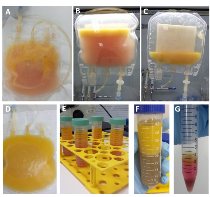
Figure 1: Steps from the protocol adipose-derived stem cells isolation. (A) Bag for lipoaspirate transport in a closed system. (B) The step of resting bag repeated three times, after washing. (C) Lipoaspirate after three washes with DPBS. (D) Lipoaspirate after collagenase digestion. (E) Lipoaspirate after digestion is distributed in a 50 mL tube. (F) Digested lipoaspirate after centrifugation. (G) Final process isolation with the pellet with the stromal vascular fraction (SVF). Please click here to view a larger version of this figure.
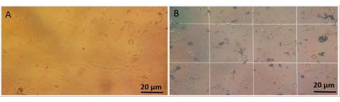
Figure 2: Morphology and viability of ADSCs. (A) Plastic adherent mesenchymal adipose-derived stem cells at the first passage after isolation at light microscopy. The cells show adhesion to the plastic and fibroblast-like morphology. (B) Trypan blue assay showing viable cells counted in Neubauer chamber using a light microscope. Please click here to view a larger version of this figure.
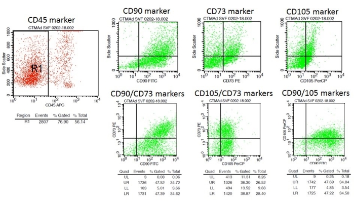
Figure 3: Subpopulation of cells positive for stem cell-associated markers in SVF of Case 9 after 8 months of cryopreservation. R1: Total cellular region analyzed in FSC (Forward Scatter) x SSC (Side Scatter) (Size x Complexity); R2: CD45 negative region, whose populations CD73, CD90, and CD105 are positive in this region. Please click here to view a larger version of this figure.
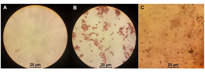
Figure 4: Differentiation assay. (A) ADSC differentiation in chondrocytes. (B) ADSC differentiation in osteocytes. (C) ADSC differentiation in adipocytes. Please click here to view a larger version of this figure.
Patient | Age at collection (years) | Weight (kg) | Height (meter) | BMI* |
Case 1 | 35 | 68 | 1.64 | 25.28 |
Case 2 | 33 | 65 | 1.65 | 23.88 |
Case 3 | 35 | 70 | 1.68 | 24.8 |
Case 4 | 34 | 72 | 1.64 | 26.77 |
Case 5 | 36 | 72 | 1.69 | 25.21 |
Case 6 | 36 | 67 | 1.64 | 24.91 |
Case 7 | 38 | 62 | 1.53 | 26.49 |
Case 8 | 50 | 63 | 1.68 | 22.32 |
Case 9 | 37 | 65 | 1.58 | 26.04 |
Table 1: Data from the samples of the individuals studied. *BMI: body mass index.
Patient | Volume collected (mL) | SVF Cellular yield (cell/mL) (x 105) | Volume in culture (mL) | Average percentage of confirmed ADSC (%) | Number of cells in initial culture (x 105) | Estimated number of ADSC in culture (x 105) | Days to P1 | Cellular yield before going to P1 (x 105) |
Case 1 | 96 | 9.2 | 2 | na | 18.4 | na | 10 | 18 |
Case 2 | 100 | 25.2 | 1 | 38 | 25.2 | 9.6 | 12 | 10.8 |
Case 3 | 100 | 26.2 | 1 | na | 26.2 | na | 12 | 6.6 |
Case 4 | 105 | 21.1 | 1 | 55.9 | 21.1 | 11.8 | 3 | 13.1 |
Case 5 | 110 | 23.7 | 1 | 61.4 | 23.7 | 14.5 | 4 | 16.1 |
Case 6 | 100 | 13.3 | 1.1 | 78.9 | 14.6 | 11.5 | 10 | 13.5 |
Case 7 | 98 | 6.8 | 2 | na | 13.6 | na | 8 | 10.5 |
Case 8 | 100 | 9.7 | 1.1 | 44.2 | 10.7 | 4.7 | 11 | 6.9 |
Case 9 | 100 | 6.1 | 2 | 43.8 | 12.2 | 5.3 | 6 | 15.9 |
SD | 3.89 | 7.81 | 0.46 | 13.75 | 5.55 | 3.53 | 3.2 | 3.79 |
Table 2: Data from different steps of the procedure from the nine patients analyzed. SVF: stromal vascular fraction; ADSC: adipose-derived stem cell; P1: passage 1; na: data not available.
SAMPLE | % Of ADSC DETERMINED BY MONOCLONAL ANTIBODIES | % OF HEMATOPOIETIC CELLS DETERMINED BY MONOCLONAL ANTIBODIES | CELL VIABILITY ASSAY AND TIME OF SVF CRYOPRESERVATION | |||||||||
CD45-(*) | CD73+/CD90+ | CD73+/CD105+ | CD105+/CD90+ | Mean | CD34+ | HLA-DR+ | CD11b+ | CD19+ | LIVE/DEAD + | |||
Case 2 | 52.34% | 31.97% | 25.36% | 56.52% | 37.95% | 63.16% | 12.87% | 2.41% | 0.21% | 39.54% (2 years) | ||
Case 4 | 48.02% | 61.62% | 40.93% | 65.25% | 55.93% | 82.94% | 26.62% | 0.00% | 0.16% | 38.30% (2 years) | ||
Case 5 | 27.74% | 54.02% | 49.72% | 80.42% | 61.38% | 73.33% | 51.31% | 0.05% | 0.00% | 23.06% (2 years) | ||
Case 6 | 55.52% | 79.52% | 67.70% | 89.52% | 78.91% | 86.86% | 8.83% | 0.18% | 1.06% | 56.76% (2 years) | ||
Case 8 | 57.28% | 46.84% | 30.88% | 57.65% | 45.12% | 78.47% | 26.97% | 0.03% | 0.00% | 55.56% (1 year) | ||
Case 9 | 56.14% | 47.52% | 36.30% | 47.69% | 43.83% | 88.10% | 26.94% | 0.05% | 0.24% | 72.34% (8 months) |
Table 3: Flow cytometry data from six of the patients. (*) From these CD45- cells, % of ADSC with different combinations of stem cell markers was determined. ADSC: adipose-derived stem cell; SVF: stromal vascular fraction; (*) From these CD45- cells, % of ADSC with different combinations of stem cell markers was determined.
Isolation yield
It is well established that the cryopreservation process, frequently required in cellular therapy, results in significant cell loss, sometimes greater than 50%29,30,35. Thus, a technical improvement for obtaining high initial cellular yield in isolation is fundamental. The collecting method of lipoaspirate and the isolation method of the cells must focus on preserving a greater number of cells, maintaining high viability, and extracting the maximum number of cells from the initial material while accounting for the long-term culture and manipulation of the cells. Therefore, straight culture maintenance is required to keep the cells away from apoptosis, senescence, or genetic instability, since cell therapy is likely to be effective and safe for patients.
To the best of our knowledge, there is no previous article using this set of steps for the isolation of mesenchymal stem cells derived from lipoaspirate, which results in a time-saving and cost-benefit technique. In this study, each methodological step was reasoned according to the literature that showed the highest final cell yield in cellular isolation. The novelty of the technique performed in this study was the use of manual aspiration associated with a series of lipoaspirate washes, with the subsequent bag resting. The collection bag used to transport and process lipoaspirate allowed undigested tissue fragments to not participate in collagenase digestion. The most critical step is adding calcium chloride to the fresh digestion solution as it potentiates the action of collagenase. The time gain is not yet reported in the literature with a cell thawing method that allows viable cells even after long cryopreservation time. The SVF cellular yield found in this study varied broadly from 6.15 to 26.2 x 10⁵ cells/mL with an average of 15.7 x 10⁵ cells/mL. This may be due to the presence of a more significant amount of adrenaline solution suctioned, which may have been higher or lower according to the stage of the surgical procedure and to the number of other known cell types generally found in the SVF. Although some studies have found negative correlations between BMI and ADSC yield36,37, this study found no significant correlation, like the other two studies38,39, decreasing the possibility of that being the cause of the incredible variety of SVF cellular yield found in this study. These data show that the lowest SVF cellular yield obtained was 6.15 x 10⁵ cells/mL. Some studies had already measured the efficiency of ADSC isolation according to the surgical technique for obtaining lipoaspirate. One study obtained 0.087 x 10⁵ cells/mL in freshly isolated SVF for the liposuction technique using adrenaline solution (as used in this study) and 0.143 x 10⁵ cells/mL without it26. This work highlighted the significance of the adrenaline solution injection due to the vasoconstrictive effect that decreases intraoperative bleeding and bruising, as the majority of surgeons choose to perform in clinical practice. Another study demonstrated that live ADSCs isolated ranged from 0 to 0.59 x 10⁵ cells/g lipoaspirate harvested, with an average of 0.295 (±0.25) x 105 cells/g tissue31. Some studies have tested different ways to achieve higher ADSC yield. One of these studies achieved about 350 x 105 for the method that presented various constituents in collagenase digestion buffer and the use of an orbital shaker40. Another study showed 29.7 (±0.2) x 105 cells/mL as the total number of SVF cells from the abdominal area41. The abdominal area chosen in this study is still the reference area for the best availability and accessibility of lipoaspirate42. The body region for lipoaspirate collection, among other factors as donor age and method of the collection chosen, is a strong determinant of the quality of the ADSC yields.
The possibility of microorganisms' contamination causing the unavailability of cells to continue the experiments was the only execution problem that limited the completion of this study. Even using antibiotics and Good Manufacturing Practice requirements, contamination can occur due to the lack of a total aseptic environment to perform the aspiration of fat.
SVF immunophenotyping
According to The Mesenchymal and Tissue Stem Cell Committee of the International Society for Cellular Therapy34, one of the three minimal criteria to define human mesenchymal stem cells is that the cells must express CD10534, CD7334, and CD9034 and should not express CD4534, CD3434, CD1434, or CD11b34, CD79a34 or CD1934, and HLA-DR34 surface membrane molecules. Mitchell43 tested fresh SVF cells by immunophenotyping and found a maximum of 54% of cells with ADSC surface markers. In this study, the immunophenotyping revealed a higher percentage of confirmed ADSC (non-hematopoietic cells) of up to 78.91% in the SVF after long-term cryopreservation (ranging from 37.95%-78.91% with a mean of 53.68%). Evidence shows that the progenitors of a stem cell population not yet committed do not express the CD34 marker34,44,45. Depending on the stage of differentiation, the CD3434 negative stem cells can generate not only hematopoietic progenitors but also more specific mesenchymal precursors, such as osteoclasts, chondrocytes, myocytes, adipocytes, and others. Some studies demonstrated the striking plasticity of the primitive stem cell population, composed of cells with stromal cell function and hematopoietic and mesenchymal progenitors45. According to the literature, the complete CD3434 functional role in the tissue formation in SVF cells is still unknown46. Mitchell43 showed a mean of 60% cells of SVF expressing CD3434 marker, whereas, in this study, the mean was 78.81%. It is known that the expression of CD3434 surface marker decreases along passages.
Adherent cells and differentiation assay
Depending on the number of cells obtained after isolation, the number of cells seeded in the first culture varied. The first culture time for reaching 80%-90% confluence in 75 cm2 flasks took an average of 8.4 days and a standard deviation of 7.5 (±4.5) days ranging from 6.6 to 16.1 x 10⁵ cells/mL. It is to be noted that even for the cases with lower cellular yield, the first culture time led to high cellular yield indexes compared to the literature, probably due to the best availability of viable cells maintained during the entire collection and isolation process. One study obtained a cellular yield of 3.75 (±1.42) x 105 ADSC per mL of lipoaspirate within a 4.1 (±0.7) day culture period47. Another study demonstrated a yield of nucleated SVF cells of 3.08 (±1.40) x 105 per millimeter with a mean of 6.0 (±2.4) days in the first culture period43. In this study, by estimating the number of ADSC seeded in culture, which varied as a function of the number of cells observed in the SVF from the collection of the same volume of 100 mL of lipoaspirate, it was verified that the number of days to reach P1 had no relation to cell volume. For example, for Case 9, in which 12.2 x 10⁵ cells were incubated, 6 days were required to reach 80%-90% confluence. For Case 6, in which 14.6 x 10⁵ cells were seeded in culture, a more extended period was necessary (10 days) to reach up to the same level of confluence. Perhaps, a minimum ADSC number is enough for the first adhesion period. There may be significant interindividual variation, such as comorbidities, age, and general health status. Some studies in the literature questioned the importance of considering the patients' inter-individual variability for SVF cell yield48,49.
According to The Mesenchymal and Tissue Stem Cell Committee of the International Society for Cellular Therapy34, mesenchymal cell must have the ability to differentiate into three different cell types as osteogenic, adipogenic and chondrogenic lineages as it was demonstrated in Figure 4.
Cell viability, cryopreservation, and genetic instability
Different methods have been used to determine viability loss, defined as plasma membrane integrity damage50. However, the cultures can also present early apoptotic cells that these approaches can ignore because they maintain an intact plasma membrane, but they are nonviable. The results reported here show a great range in viability marker, from 23.06%-72.34%, with a mean of 47.6% after long-term cryopreservation. Considering that cryopreservation is a significant step concerning cell therapy, recovery of the maximum number of viable and functional stem cells after thawing is one of the priority issues for the success of cell therapy. The literature has shown that at least 50% of cell viability is lost from 1-4 months after cryopreservation43. Notably, the lowest indexes presented in this study are from Case 5, Case 4, and Case 2, which are the oldest cryopreserved samples (about 2 years). Although they presented the lowest viability indexes, they demonstrated high cellular yield in trypan blue dye exclusion assay in fresh SVF. Although the literature supports causes of viability loss, these rates are lower than expected. Temperature fluctuations in cell storage due to technical reasons can cause the increase and accumulation of stress and favor the accumulation of aqueous portions, generating crystals that damage the plasmatic membrane during long-term cryopreservation51. The literature shows more than 70% viability for samples with the same or longer freezing time. However, the viability check was performed with a different technique than that carried out in this study52. Another study showed that longer cryopreservation negatively affects cellular viability31, which can be explained by temperature variations in the -80 °C freezer. Hence, cells often need to stay too long in culture, which increases cell cycle stress, bringing risks for genetic instability and consequently compromising cellular therapy. There is also a consensus in the literature indicating some stress factors and how they affect cell cytogenetic stability, which is essential to maintain the prolonged stem cell cultivation required for cell therapy35,53.
Methodology benefit
To date, literature shows no standardized protocol to isolate ADSC aiming for clinical applications. Most of the studies demonstrate complex, time-consuming protocols24. In this study, the efficiency of the method versus the time demanded to complete the initial cellular yield must be emphasized: about 1.5 h. According to literature, isolation of adipose-derived stem cells can take about 3 h to 8 h54,55. Thus, the gain of time allied to the high cellular income is critical for regenerative medicine therapeutics advancement. More cell viability assessments should be carried out parallel to those performed in this work to improve this method. Further randomized controlled trials incorporating a more extensive sample using this methodology are required to countersign these results.
The authors have nothing to disclose.
We thank the patients who volunteered to participate and the medical and nursing staff of the Hospital São Paulo. This study was supported by the Fundação de Amparo à Pesquisa do Estado de São Paulo (FAPESP) and Conselho Nacional de Desenvolvimento Científico e Tecnológico (CNPq), Brazil.
1.8 mL cryovials | Nunc Thermo Fisher Scientific | 340711 | |
150 mL polyvinyl chloride transfer bag | JP FARMA | 80146150059 | |
2% Alizarin Red S Solution, pH 4.2 | Sigma Aldrich | A5533 | |
Adrenaline (1 mg/mL) | Hipolabor | NA | |
Alcian Blue solution | Sigma Aldrich | 1,01,647 | |
Antibiotic-Antimycotic 100x | Gibco | 15240062 | |
BD FACSCalibur Flow Cytometer using BD CellQues Pro Analysis | BD BioSciences | NA | |
Calcium chloride 10% | Merck | 102379 | |
Chlorhexidine gluconate 4% | VIC PHARMA | NA | |
Collagenase, Type I, powder | Gibco | 17018029 | |
DMEM (Dulbecco's modified Eagle's medium) | Gibco | 11966025 | |
DPBS no calcium, no magnesium (Dulbecco's Phosphate Buffered Saline Gibco Cell Therapy Systems) | Gibco | A1285801 | |
DPBS with calcium (Dulbecco's Phosphate Buffered Saline Gibco Cell Therapy Systems) | Gibco | A1285601 | |
Fetal bovine serum | Gibco | 10500056 | |
Formaldehyde 4% | Sigma Aldrich | 1,00,496 | |
Inverted light microscope | Nikon Eclipse TS100 | NA | |
Live and Dead Cell Assay | Thermofisher | 01-3333-41 | 01-3333-42 | |
Monoclonal antibody: CD105 | BD BioSciences | 745927 | |
Monoclonal antibody: CD11B | BD BioSciences | 746004 | |
Monoclonal antibody: CD19 | BD BioSciences | 745907 | |
Monoclonal antibody: CD34 | BD BioSciences | 747822 | |
Monoclonal antibody: CD45 | DAKO | M0701 | |
Monoclonal antibody: CD73 | BD BioSciences | 746000 | |
Monoclonal antibody: CD90 | BD BioSciences | 553011 | |
Monoclonal antibody: HLA-DR | BD BioSciences | 340827 | |
Mr. Frosty Freezing Container | Thermo Fisher Scientific | 5100-0001 | |
PBS (phosphate buffered saline) 1x pH 7.4 | Gibco | 10010023 | |
StemPro Adipogenesis Differentiation Kit | Gibco | A1007001 | |
StemPro Chondrogenesis Differentiation Kit | Gibco | A1007101 | |
StemPro Osteogenesis Differentiation Kit | Gibco | A1007201 | |
Sterile connector with one spike with needle injection site | Origen Biomedical Connector, USA | NA | Code mark: IBS |
Trypan blue solution 0.4% | Sigma Aldrich | 93595 | |
Trypsin-EDTA 0.25% 1x, phenol red | Gibco | 25200056 |