Decellularized extracellular matrix (dECM) can provide suitable microenvironmental cues to recapitulate the inherent functions of target tissues in an engineered construct. This article elucidates the protocols for the decellularization of pancreatic tissue, evaluation of pancreatic tissue-derived dECM bioink, and generation of 3D pancreatic tissue constructs using a bioprinting technique.
The transplantation of pancreatic islets is a promising treatment for patients who suffer from type 1 diabetes accompanied by hypoglycemia and secondary complications. However, islet transplantation still has several limitations such as the low viability of transplanted islets due to poor islet engraftment and hostile environments. In addition, the insulin-producing cells differentiated from human pluripotent stem cells have limited ability to secrete sufficient hormones that can regulate the blood glucose level; therefore, improving the maturation by culturing cells with proper microenvironmental cues is strongly required. In this article, we elucidate protocols for preparing a pancreatic tissue-derived decellularized extracellular matrix (pdECM) bioink to provide a beneficial microenvironment that can increase glucose sensitivity of pancreatic islets, followed by describing the processes for generating 3D pancreatic tissue constructs using a microextrusion-based bioprinting technique.
Recently, pancreatic islet transplantation has been considered a promising treatment for patients with type 1 diabetes. The relative safety and minimal invasiveness of the procedure are great advantages of this treatment1. However, it has several limitations such as the low success rate of isolating islets and the side effects of immunosuppressive drugs. Furthermore, the number of engrafted islets decreases steadily after transplantation due to the hostile environment2. Various biocompatible materials such as alginate, collagen, poly(lactic-co-glycolic acid) (PLGA) or polyethylene glycol (PEG) have been applied to pancreatic islet transplantation to overcome these difficulties.
3D cell printing technology is emerging in tissue engineering due to its great potential and high performance. Needless to say, bioinks are known as important components for providing a suitable microenvironment and enabling the improvement of cellular processes in printed tissue constructs. A substantial number of shear-thinning hydrogels such as fibrin, alginate, and collagen are widely used as bioinks. However, these materials show a lack of structural, chemical, biological, and mechanical complexity compared to the extracellular matrix (ECM) in native tissue3. Microenvironmental cues such as the interactions between islets and ECM are important signals for enhancing the function of islets. Decellularized ECM (dECM) can recreate the tissue-specific composition of various ECM components including collagen, glycosaminoglycans (GAGs), and glycoproteins. For example, primary islets that retain their peripheral ECMs (e.g., type I, III, IV, V, and VI collagen, laminin, and fibronectin) exhibit low apoptosis and better insulin sensitivity, thus indicating that tissue-specific cell-matrix interactions are important for enhancing their ability to function similarly to original tissue4.
In this paper, we elucidate protocols for preparing pancreatic tissue-derived decellularized extracellular matrix (pdECM) bioink to provide beneficial microenvironmental cues for boosting the activity and functions of pancreatic islets, followed by the processes for generating 3D pancreatic tissue constructs using a microextrusion-based bioprinting technique (Figure 1).
Porcine pancreatic tissues were collected from a local slaughterhouse. Animal experiments were approved by the Institutional Animal Care and Use Committee (IACUC) of Asan Medical Center, Seoul, Korea.
1. Tissue decellularization
2. Assessment of decellularized tissues
NOTE: To evaluate the residual amount of dsDNA, glycosaminoglycans (GAGs), and collagen in the decellularized tissue compared to native tissue, at least 1 g of each of the non-decellularized tissue (native tissue) and decellularized tissue are required for one batch of assessment. The amount of dsDNA, GAGs, and collagen can be calculated based on the dry weight of the tissue.
3. Bioink preparation
NOTE: pdECM powder can be stored stably at -80 °C for at least one year. Before pH adjustment, the digested pdECM solution can be stored at -20 °C for one month. Prior to use, thaw the sample of frozen pdECM solution at 4 °C overnight. The pH-adjusted pdECM solution can be stored at 4 °C for up to one week. The digested pdECM solution can be stored at 4 °C for at least a few days but should not exceed 1 week.
4. Rheological analysis
5. 3D cell printing of pancreatic tissue constructs using islet
6. 3D cell printing of pancreatic construct with patterned structure
Decellularization of pancreatic tissues
We developed the process for preparing pdECM bioink to provide pancreatic tissue-specific microenvironments for enhancing functionality of islets in a 3D bioprinted tissue construct (Figure 2A). After the decellularization process, 97.3% of dsDNA was removed and representative ECM components such as collagen and GAGs remained at 1278.1% and 96.9% compared to that of the native pancreatic tissue, respectively (Figure 2B).
Bioink preparation
To apply the pdECM in the printing process, the pdECM powder was solubilized in weak acid with pepsin and neutralized using 10 M NaOH solution. The digested pdECM solution could then be diluted through mixing with a cell culture medium or 1x PBS. In this study, we prepared pdECM bioink at a final concentration of 1.5% for further study. The pdECM bioink maintained a solution phase when it was placed under room temperature and instantly converted into a gel phase after incubation at 37 °C for 30 min. To investigate the effect of the pdECM bioink on islets, isolated islets were encapsulated in the pdECM, alginate and collagen bioinks at a concentration of 1.5%. The result of the glucose-stimulated insulin secretion test showed islets in the pdECM bioink represented the highest index (approximately 3.174) among the experimental groups, indicating higher functionality over the widely applied hydrogels for islet encapsulation5.
Rheological analysis
Viscosity is one of the critical characteristics when considering a printable biomaterial. We measured viscosity of the pdECM bioink at a frequency ranging from 1 to 1,000 Hz at 15 °C for printing various dECM bioinks6,7,8. The pdECM bioink showed shear-thinning behavior and the value was approximately 10 Pa·s at the shear rate of 1/s, indicating the pdECM bioink had appropriate rheological characteristics for extrusion through a nozzle (Figure 3A). The gelation kinetics at a temperature ranging from 4 to 37 °C indicated the gelation behavior of the pdECM bioink at physiologically relevant temperatures. The complex modulus started to increase when the temperature reached 15 °C, and it increased rapidly when the temperature was maintained at 37 °C, indicating the sol-gel transition of the pdECM bioink (Figure 3B). The dynamic G' and G" of pdECM bioink were investigated at physiologically relevant temperatures to ensure its stability after the printing process, which resulted in having a stable modulus under the frequency sweep condition (Figure 3C).
3D cell printing
3D cell-laden pancreatic tissue constructs were fabricated by using a microextrusion-based printing process. To build a construct containing at least 3,000 Islet equivalents (IEQ), that corresponds to the tissue volume of a perfectly spherical islet with a diameter of 150 µm9, we designed the construct with a dimension of 10 mm x 10 mm x 3 mm (Figure 4A). The process parameters and conditions for printing pancreatic islets were selected to encapsulate islets, which are large cellular clusters in sizes ranging 100-250 µm in diameter (Figure 4B). Using a multi-head printing system, various types of 3D constructs-such as the shape of the lattice having alternate lines of blue and red-were fabricated by using the developed pdECM (Figure 4C), indicating the versatility of pdECM for the purpose of 3D bioprinting to harmonize two or more types of living cells in a tissue-like arrangement.
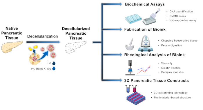
Figure 1: Schematic of the development of decellularized pancreatic tissue, evaluation of pdECM bioink and fabrication of 3D pancreatic tissue constructs. Please click here to view a larger version of this figure.
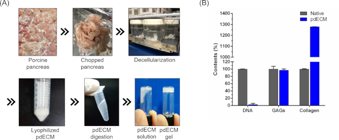
Figure 2: Representative images of the decellularization process and biochemical characterization of pdECM. (A) Overview of the decellularization of porcine pancreatic tissue. (B) Results of biochemical assays of native tissue and pdECM. Error bars show standard deviation. Copyright (2019) The Royal Society of Chemistry5. Please click here to view a larger version of this figure.
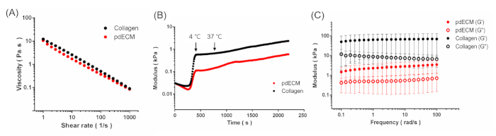
Figure 3: Rheological analysis of pdECM bioink. (A) Viscosity of pdECM and collagen bioinks that exhibited shear thinning behavior. (B) Gelation kinetics of pdECM and collagen bioinks during temperature change. (C) The complex modulus of crosslinked pdECM and collagen bioinks. Copyright (2019) of The Royal Society of Chemistry5. Please click here to view a larger version of this figure.
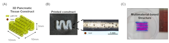
Figure 4: 3D cell printing of cell-laden pdECM bioink for 3D pancreatic tissue constructs. (A) The dimensions of 3D pancreatic tissue constructs. (B) Pancreatic islet-laden and (C) multimaterial-based 3D pancreatic tissue constructs. Copyright (2019) of The Royal Society of Chemistry5. Please click here to view a larger version of this figure.
This protocol described the development of pdECM bioinks and the fabrication of 3D pancreatic tissue constructs by using 3D cell printing techniques. To recapitulate the microenvironment of the target tissue in the 3D engineered tissue construct, the choice of bioink is critical. In a previous study, we validated that tissue-specific dECM bioinks are beneficial to promote stem cell differentiation and proliferation10. Compared to synthetic polymers, dECM can serve as a cell-favorable environment because of the tissue-specific composition and architecture11. Therefore, the decellularization process should be seriously considered for the high retention of major components in the dECM.
The selection of different detergents for decellularization of pancreatic tissue varies the residual ECM constituent12. In the process of decellularization, we noticed that the use of sodium dodecyl sulfate (SDS) can affect loss of the ECM proteins13. Thus, we modified our previous protocol by eliminating the step for the treatment of SDS solution, which is an ionic surfactant used in many cleaning and decellularization processes featuring relatively harsh characteristics compared to the others such as Trion-X 100, or 3-[(3-cholamidopropyl) dimethyl-lammonio]-1-propanesulfonate (CHAPS). In this protocol, we used 1% Triton-X 100 solution for 84 h instead of SDS solution, which was able to remove the cellular components effectively while preserving GAGs and collagenous proteins. In addition, we noted that removal of residual lipids by treating with IPA is also a very crucial process for inducing the crosslinking of pdECM bioink and it can be understood in the same context as a previously published article4. Treatment with peracetic acid solution was also applied for the sterilization of decellularized tissue. In addition, removal of the remaining detergents and chemicals in the decellularized tissue is a crucial step to prevent the inflammatory host response. However, we did not discuss that issue in this protocol. Protocols that include a sanitization process at the end of decellularization will improve the biocompatibility of the decellularized material. Furthermore, standards for evaluation criteria should be considered to ensure that detergents and chemicals are completely removed.
The digestion of pdECM bioink with pepsin was performed to achieve the homogenous mixing of pdECM powder in the acidic solution by cleavage of the telopeptide region in the collagenous protein. In the pH adjustment process, keeping the pdECM bioink on ice is critical for the preservation of gelation. Afterward, we can produce physically crosslinkable pdECM pre-gel bioinks that can enter a gel state by incubating at 37 °C, which is one of the main advantages of dECM-based bioinks. Selection of the proper concentration of the pdECM bioink is also important10. An ideal bioink should protect cells from external damage that occurs during the printing process such as pneumatic pressure and temperature change. It is known that the applied shear force may cause damage to the cells and reduce the cell viability in the printed constructs10. Also, enhancing concentrations of bioink could induce cell death5. In contrast, low concentrations of bioink induces low viscosity which means poor printability and shape-fidelity during printing. It is necessary to check the viscosity of bioink and optimize its concentration.
Currently, researchers are actively studying the development of various types of tissue-derived bioinks for printing 3D tissue constructs14,15,16. The results of these studies indicate that the bioink could provide tissue-specific microenvironments for cells. These unique conditions can promote the differentiation or maturation of stem cells and the proliferation of cells. Moreover, utilizing the multi-head equipped 3D cell-printing system makes it possible to print multiple types of bioinks with high precision simultaneously. Using this technique, a structure with a specific pattern can be produced, thus showing design versatility. In addition, it is feasible to encapsulate different types of cells into each bioink to mimic native cell arrangement17. These patterned structures can be utilized in the induction of vascularization or co-culture effect by improving cell-to-cell interactions, which can be key factors in the long-term survival of specific cells18,19.
The authors have nothing to disclose.
This research was supported by the Bio & Medical Technology Development program of the National Research Foundation (NRF) funded by the Korean government (MSIT) (2017M3A9C6032067) and "ICT Consilience Creative Program" (IITP-2019-2011-1-00783) supervised by the IITP (Institute for Information & Communications Technology Planning & Evaluation).
Biological Safety Cabinets | CRYSTE | PURICUBE 1200 | |
Deep Freezer | Thermo Scientific Forma | 957 | |
Digital orbital shaker | DAIHAN Scientific | DH.WSO04010 | |
Dry oven | DAIHAN Scientific | WON-155 | |
Freeze dryer | LABCONCO | 7670540 | |
Fridge | SANSUNG | CRFD-1141 | |
Grater | ABM | 1415605793 | |
Inverted Microscopes | Leica | DMi1 | |
Microcentrifuge | CRYSTE | PURISPIN 17R | |
Microplate reader | Thermo Fisher Scientific | Multiskan GO | |
Mini centrifuge | DAIHAN Scientific | CF-5 | |
Multi-Hotplate Stirrers | DAIHAN Scientific | SMHS-6 | |
Nanodrop | Thermo Fisher Scientific | ND-LITE-PR | |
pH benchtop meter | Thermo Fisher Scientific | STARA2110 | |
Rheometer | TA Instrument | Discovery HR-2 | |
Vortex Mixer | DAIHAN Scientific | VM-10 | |
Cirurgical Instruments | |||
Operating Scissors | Hirose | HC.13-122 | |
Forcep | Korea Ace Scientific | HC.203-30 | |
Materials | |||
1.7 mL microcentrifuge tube | Axygen | MCT-175-C | |
10 ml glass vial | Scilab | SL.VI1243 | |
40 µm cell strainer | Falcon | 352340 | |
5 L beaker | Dong Sung Science | SDS 2400 | |
50 mL cornical tube | Falcon | 352070 | |
500 mL beaker | Korea Ace Scientific | KA.23-08 | |
500 mL bottle-top vacuum filter | Corning | 431118 | |
500 mL plastic container | LOCK&LOCK | INL301 | |
96well plate | Falcon | 353072 | |
Aluminum foil | DAEKYO | ||
Kimwipe | Kimtech | ||
Magnetic bar | Korea Ace Scientific | BA.37110-0003 | |
Mortar and pestle | DAIHAN Scientific | SC.MG100 | |
Multi-channel pipettor | Eppendorf | 4982000314 | |
Petri Dish | SPL | 10100 | |
pH indicator strips | Sigma-Aldrich | 1095350001 | |
Sieve filter mesh | DAIHAN Scientific | ||
Decellularization | |||
10x pbs | Hyclone | SH30258.01 | |
4.7% Peracetic acid | Omegafarm | ||
70% ethanol | SAMCHUN CHEMICALS | E0220 SAM | |
Distilled water | |||
IPA | SAMCHUN CHEMICALS | samchun I0348 | |
Triton-X 100 | Biosesang | T1020 | |
Biochemical assay | |||
1,9-Dimethyl-Methylene Blue zinc chloride double salt | Sigma-Aldrich | 341088 | |
10 N NaOH | Biosesang | S2018 | |
Chloramine T | Sigma-Aldrich | 857319 | |
Chondroitin sulfate A | Sigma-Aldrich | C4384 | |
Citric acid | Supelco | 46933 | |
Cysteine-HCl | Sigma-Aldrich | C1276 | |
Glacial acetic acid | Merok | 100063 | |
Glycine | Sigma-Aldrich | 410225 | |
HCl | Sigma-Aldrich | H1758 | |
Na2-EDTA | Sigma-Aldrich | E5134 | |
NaCl | SAMCHUN CHEMICALS | S2097 | |
Papain | Sigma-Aldrich | p4762 | |
P-DAB | Sigma-Aldrich | D2004 | |
Perchloric acid | Sigma-Aldrich | 311421 | |
Sodium acetate | Sigma-Aldrich | S5636 | |
Sodium hydroxide | Supelco | SX0607N | |
Sodium phosphate(monobasic) | Sigma-Aldrich | RDD007 | |
Toluene | Sigma-Aldrich | 244511 | |
Bioink | |||
Charicterized FBS | Hyclone | SH30084.03 | |
Penicillin-Streptomycin | Thermo Fisher Scientific | 15140122 | |
Pepsin | Sigma-Aldrich | P7215 | |
Rose bengal | Sigma-Aldrich | 198250 | |
RPMI-1640 medium | Thermo Fisher Scientific | 11875093 | |
Trypan Blue solution | Sigma-Aldrich | T8154 |